
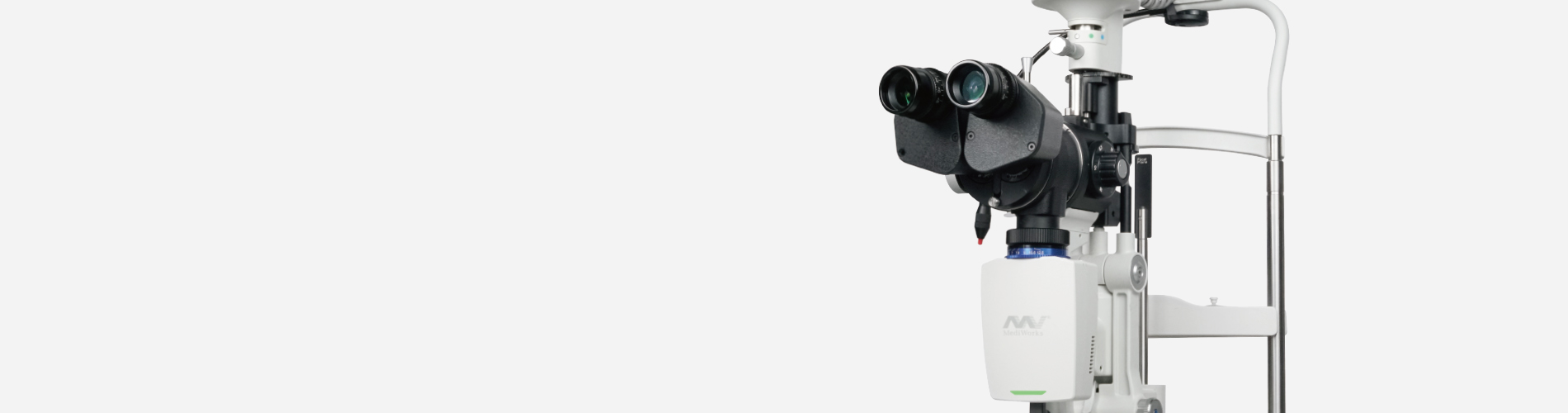
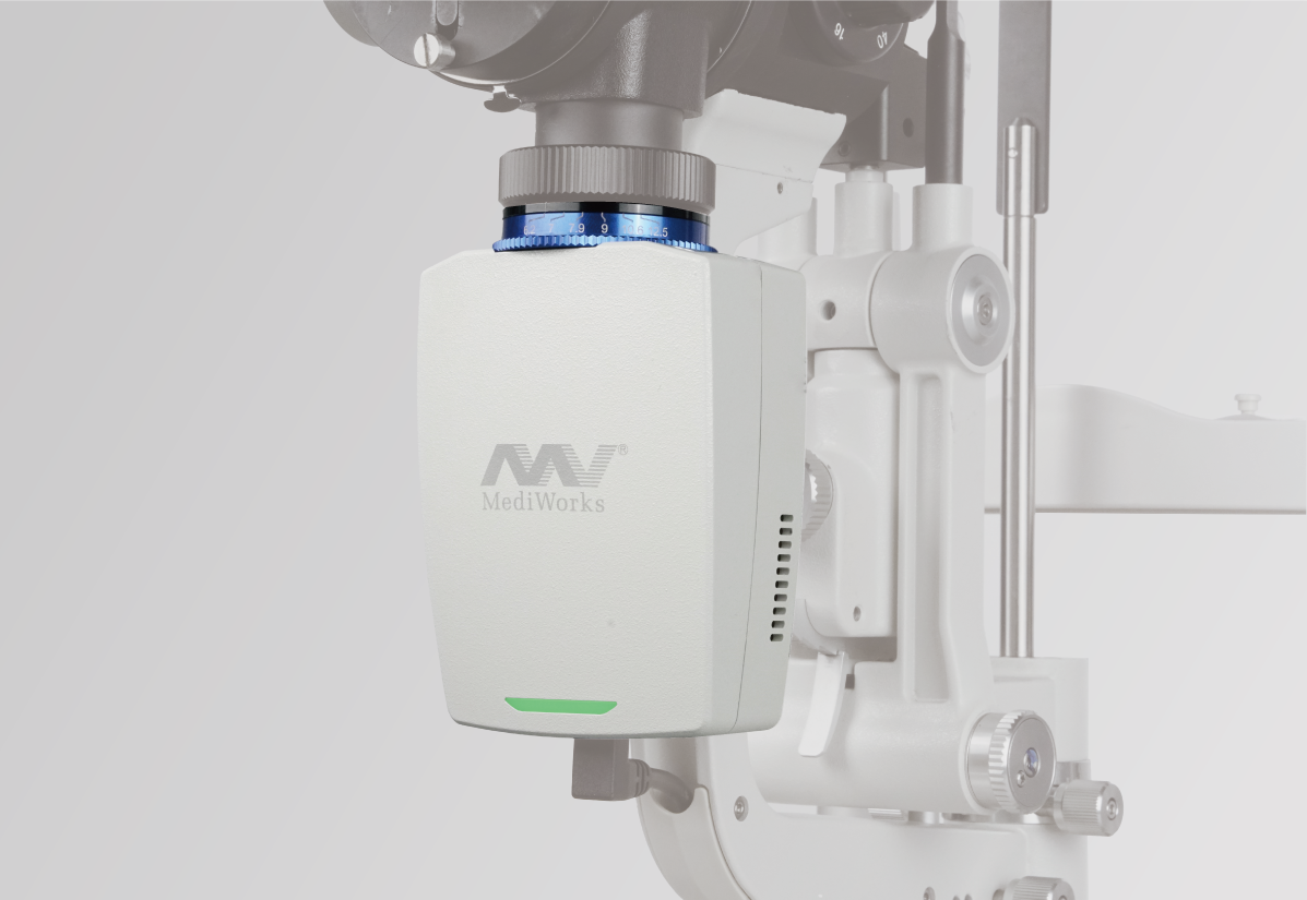
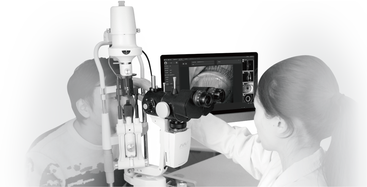
The design of the ophthalmic slit lamp S390L(Firefly) was inspired by the shape of the firefly. The smart design largely saves space for clinicians compared to other bulky camera systems. We have preset many camera parameters so the user does not need to adjust settings before using the device. The user can operate the machine immediately once the installation has been finished. The device has the following automatic functions for photo shooting and processing when equipped with our Mediview software:
Wide Dynamic Range
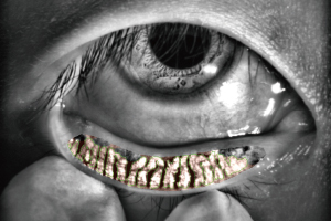
Meibomian Glands Examination
Auto Exposure
Auto Gain
Auto White Balance
Auto OD/OS Indicator
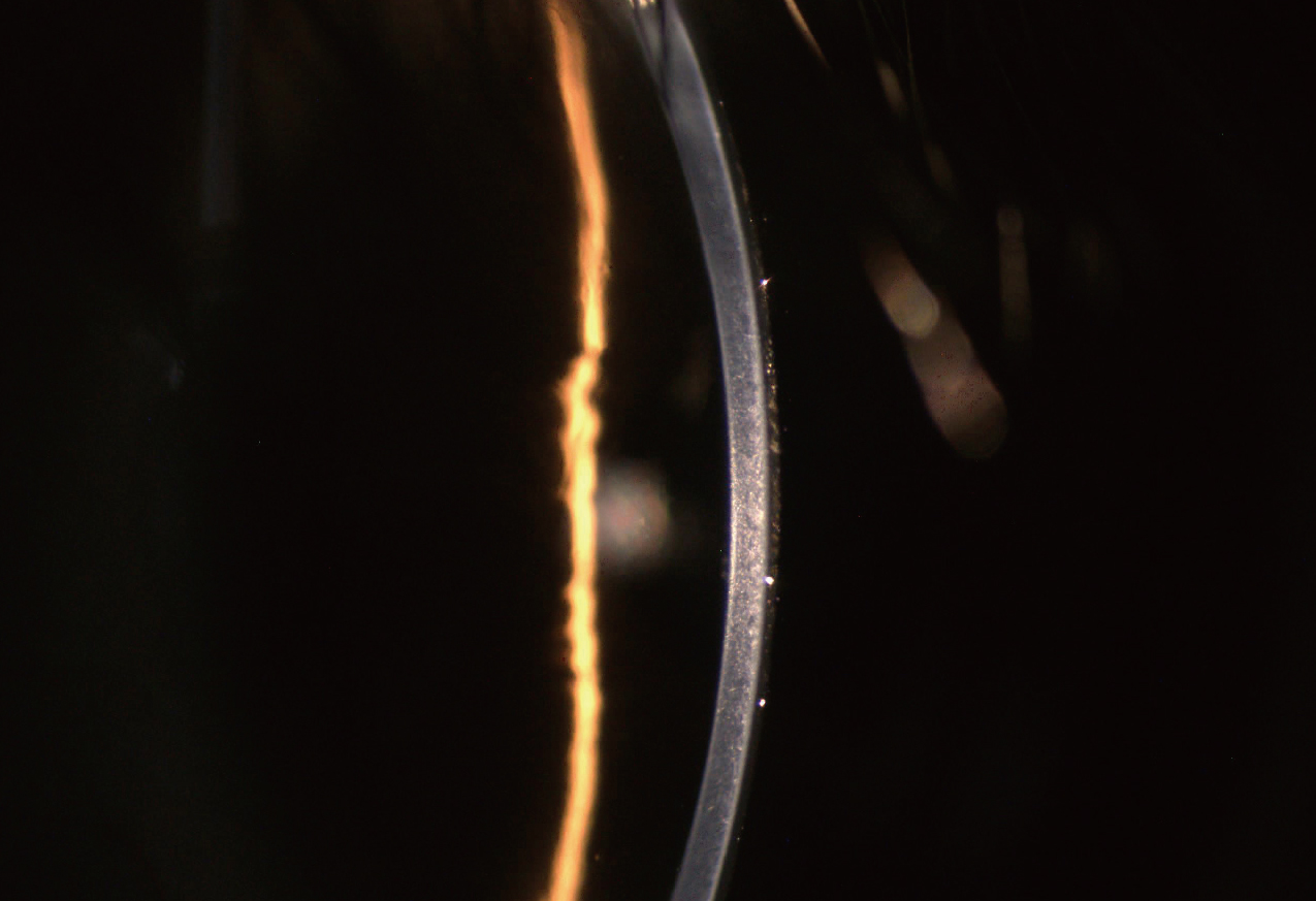
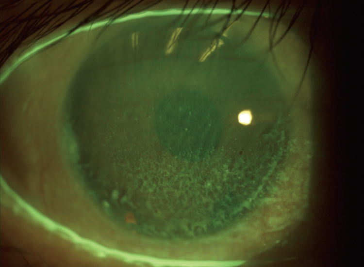
The slit is still clear and sharp under weak light.
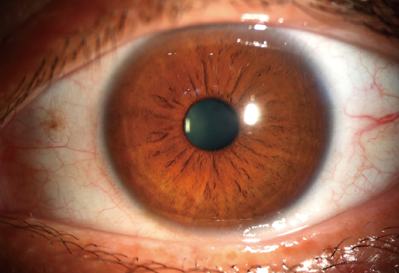
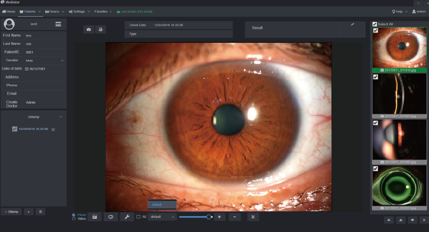
Iris and sclera images are simultaneously clearly presented with more realistic and evenly distributed color.
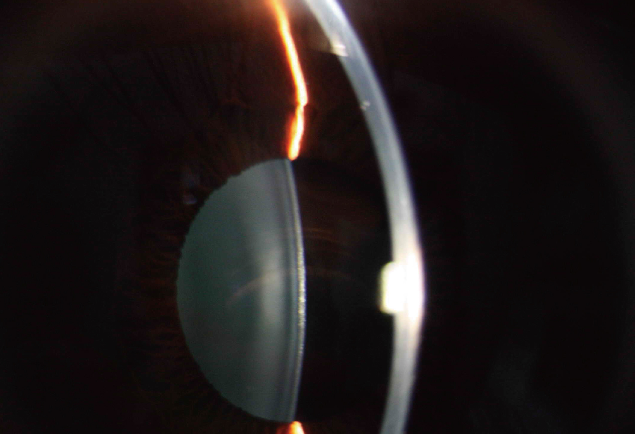
Optical resolution is up to 2700·N lp/mm
(200 lp/mm), providing more details of the pathologies.
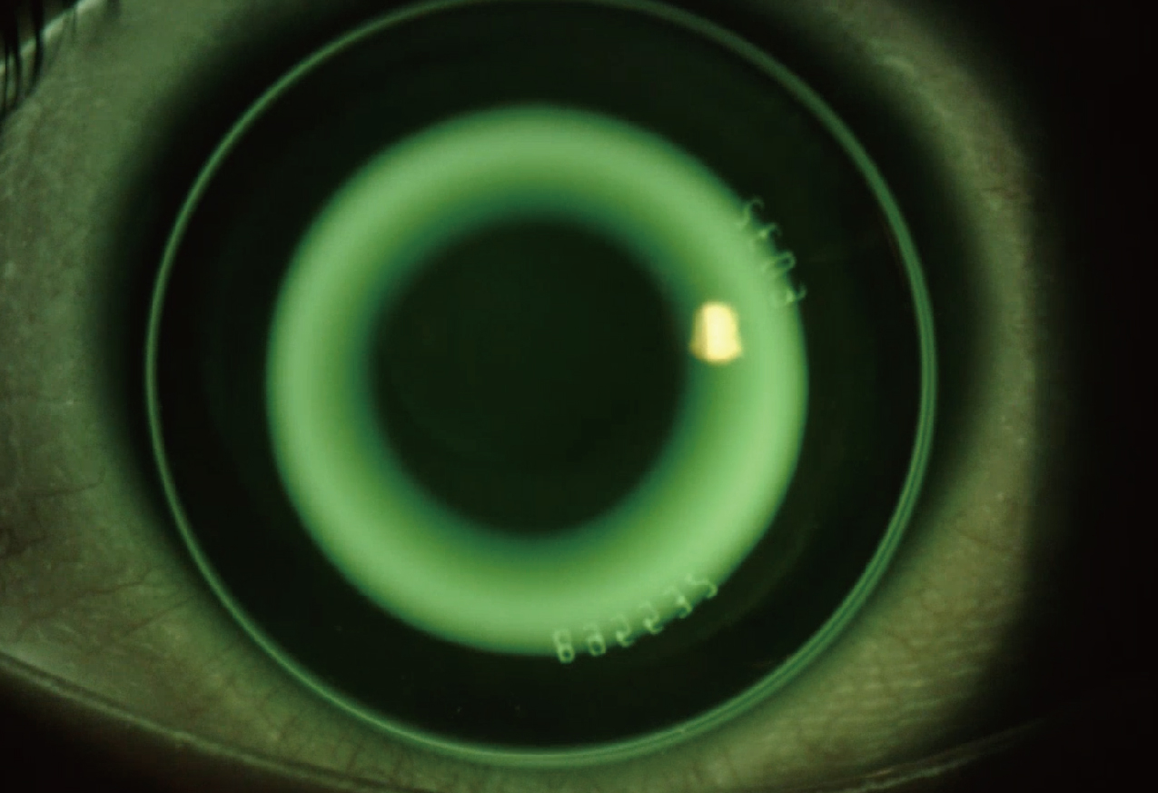
Built-in yellow filter along with cobalt-blue filter increases the contrast of Sodium Fluorescein Staining image.Increase positive rate of early corneal epithelial staining.
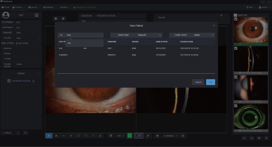
The patient management system enables clinicians to build and edit patient record,search information by inputting keywords.Clinicians can easily record symptoms and manage the data all the time. The software supports DICOM which makes the images captured by Firefly be easily integrated into hospital's medical system.
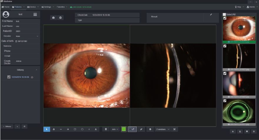
Clinicians can measure the pathology area with our powerful software tools and change the contrast and brightness of the images. Clinicians can also compare several images at one time to analyze the symptoms and pathology.
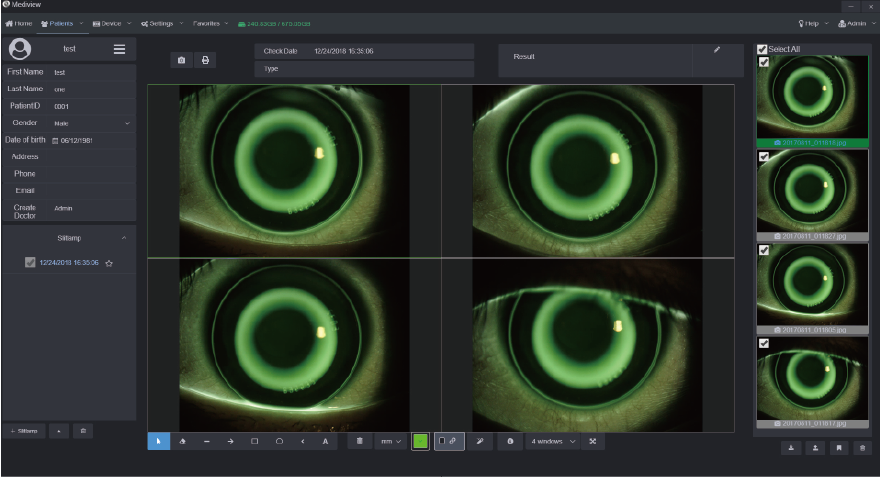
The optometrists can capture and record high resolution fluorescein images of lens fitting and real-time video without a recording time limit. By comparing the different lens fitting effects, the optometrist can show and educate patients which lens is most suitable for them.
Clinicians can customize auto exposure values according to the image demand and save as templates for future capturing purpose.
Also, the printing report can be customized according to clinician's needs.
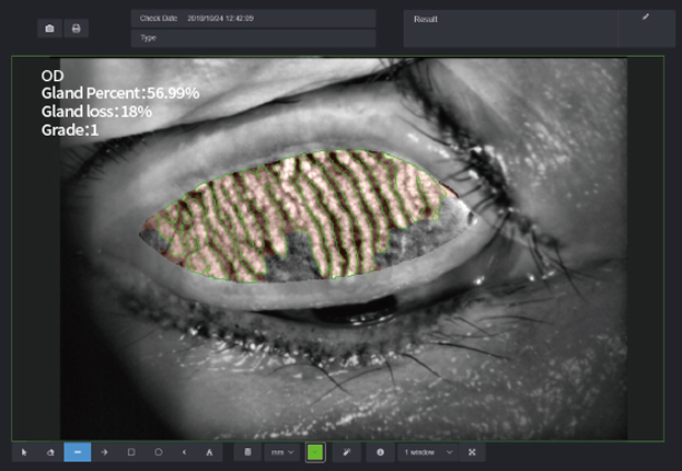
Built-in infrared light source allows the doctor to accurately judge the absence of the meibomian glands.
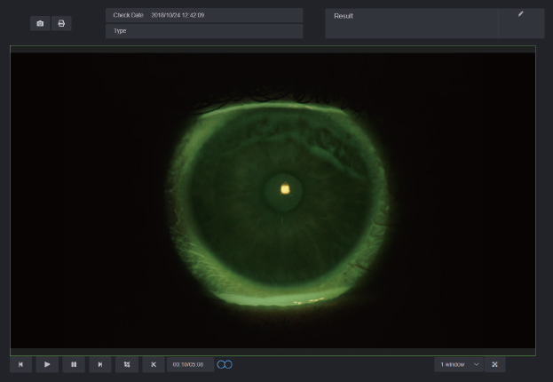
High-performance digital module, doctors can get the tear film breakup time and judge the stability of it by high-resolution video recording.
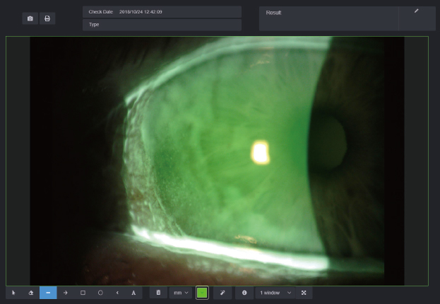
With a built-in yellow filter, doctors can accurately analyze eye surface damage and inflammation images.
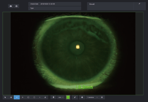
Doctors can obtain tear meniscus height by using measuring function in the Mediview software, and effectively evaluate tear
meniscus height.
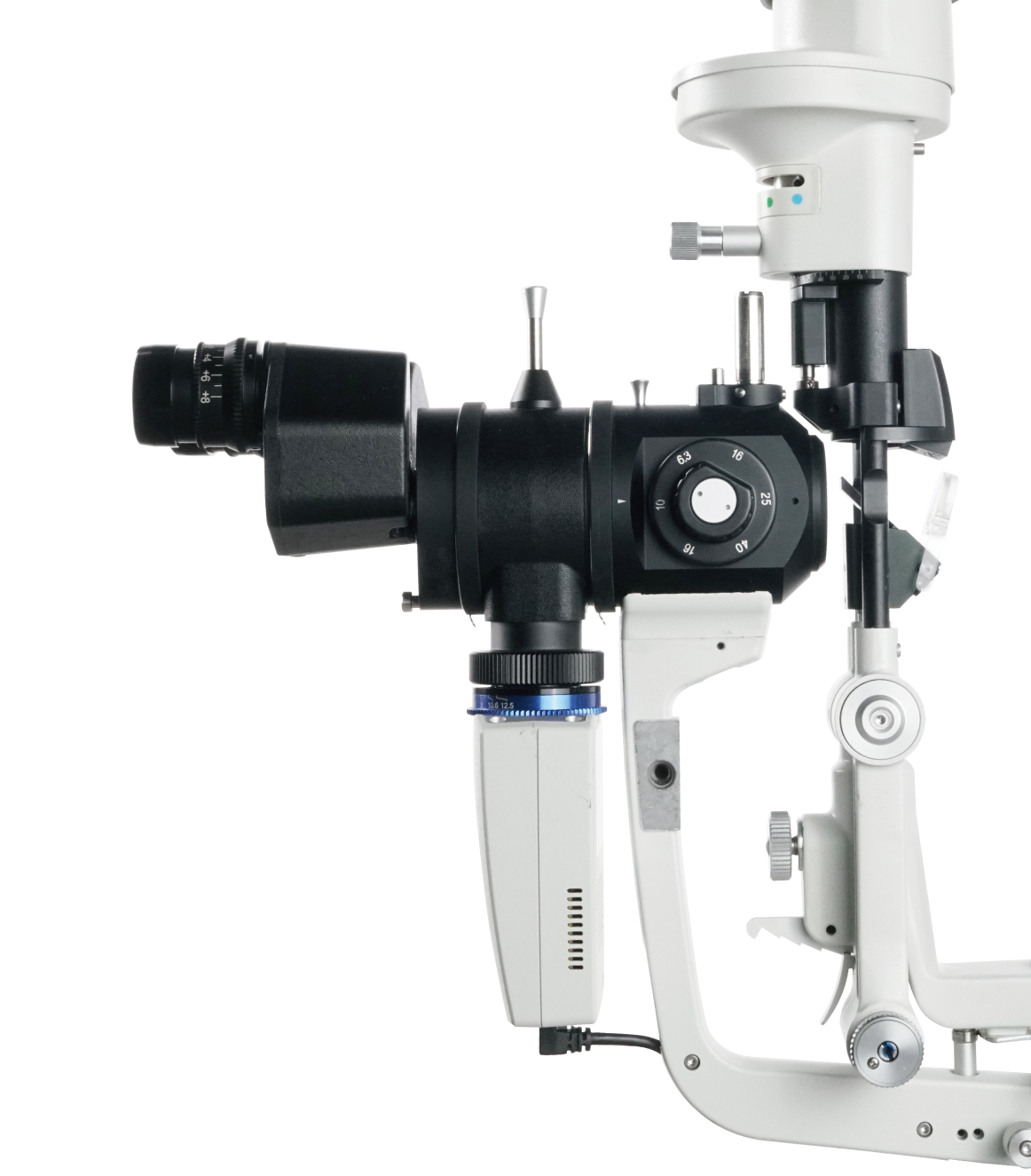
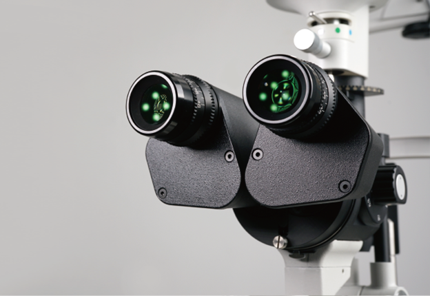
| Microscope | |
|---|---|
| Microscope Type | Galilean Type |
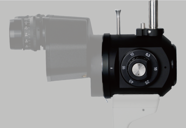
| Magnification Change | Revolving Drum 5 steps |
| Total Magnification | 6.3 x, 10 x, 16 x, 25 x, 40 x |
| Optical Resolution | 2700·N lp/mm (200 lp/mm) |
| Eyepieces | 12.5 x |
| Angle between Eyepieces | 10° |
| Pupillary Adjustment | 52 mm ~ 80 mm |
| Diopter Adjustment | - 8 D ~ + 8 D |
| Field of View | Ø36.2 mm, Ø22.3 mm, Ø14 mm, Ø8.9 mm, Ø5.7 mm |
| Slit Illumination | |
|---|---|
| Slit Width | 0 ~ 14 mm continuous (slit becomes a circle at 14 mm) |
| Slit Length | 1 ~ 14 mm continuous |
| Aperture Diameters | Ø14 mm, Ø10 mm, Ø5 mm, Ø3 mm, Ø2 mm, Ø1 mm, Ø0.2 mm |
| Slit Angle | 0°~180° |
| Slit Inclination | 5°, 10°, 15°, 20° |
| Filters | Heat-absorbing filter, ND filter, Red-free filter, Cobalt Blue filter,Yellow filter built-in |
| Lamp | LED |
| Luminance | ≥ 150 klx |
| Power Supply | |
|---|---|
| Input Voltage | ~ 100 V ~ 240 V |
| Input Frequency | 50 Hz / 60 Hz |
| Rated current | 1.2 A |
| Output Voltage | LED 3 V, Fixation 15 V |
| Packaging | |
|---|---|
| Dimension | 740 mm x 450 mm x 530 mm(L/W/H) |
| Gross weight | 23 kg |
| Net weight | 17 kg |
| System Specifications | |
|---|---|
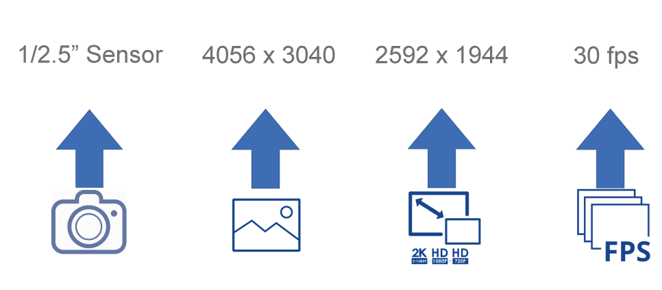
| Image Sensor | 12 M Pixels |
| Photo Resolution | 4056 x 3040 |
| Format | JPEG |
| Video Resolution | 2592 x 1944 |
| Frame of Video | 30 fps |
| Video Formats | MP4 H.264 |
| Exposure Mode | Automatic exposure |
| Transmission Interface | USB |
| Computer Specifications | |
|---|---|
| PC configuration | i5 - 10500T 8GB memory 256GB SSD + 1TB storage |
| Display | 1920 x 1080 23.8 inch |
| PC system | Windows 10 |