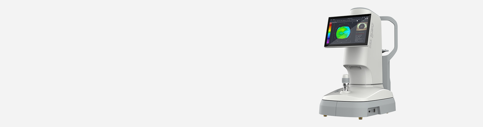
DEA 520 is a multi-purpose corneal topographer that integrated dry eye and corneal topography analysis.
Thousands of measure points - ensure more data available and accurate analysis
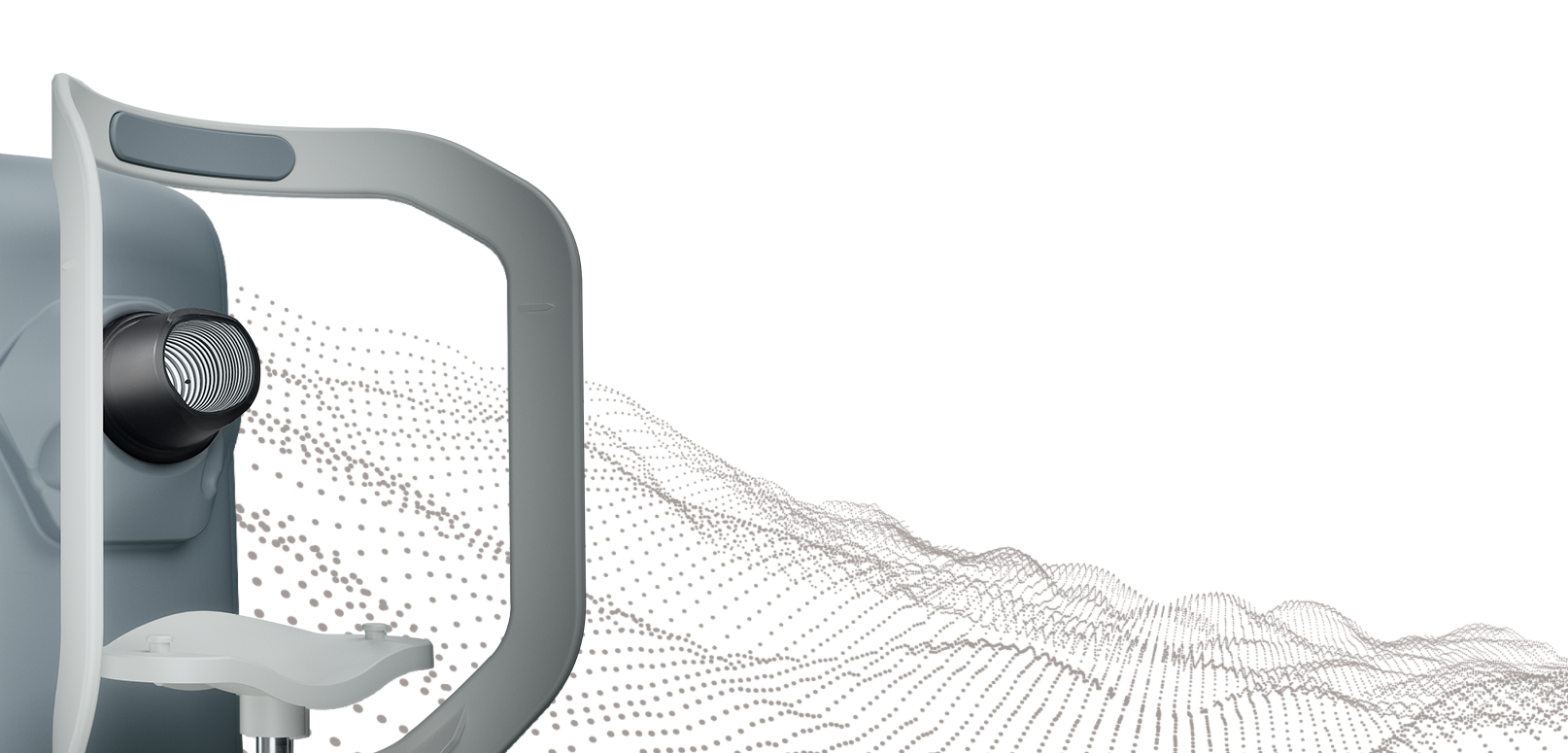
Smaller cone design - bigger projection area
3 Illuminations - white illumination, infrared illumination,cobalt blue illumination
● Dry Eye Questionnaire
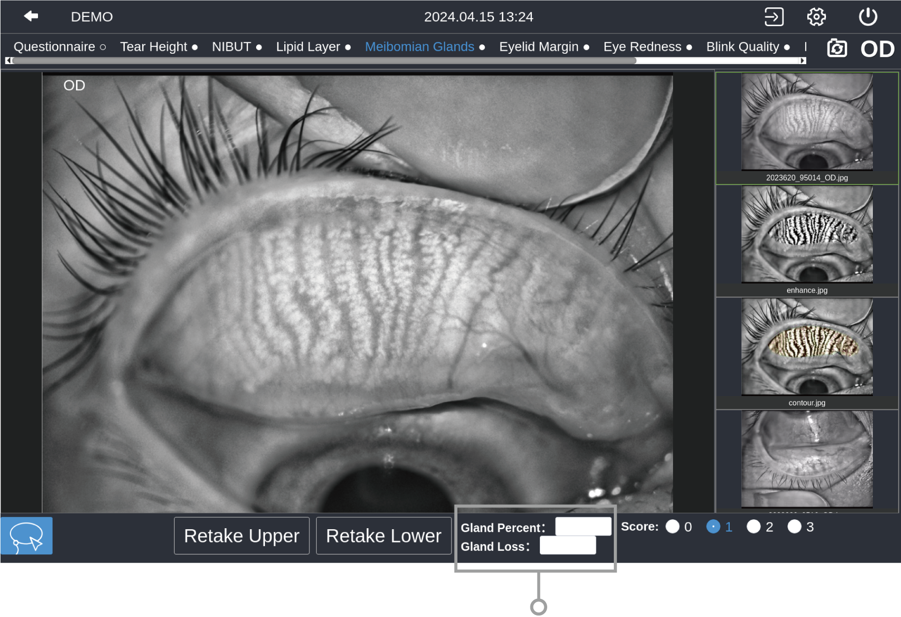
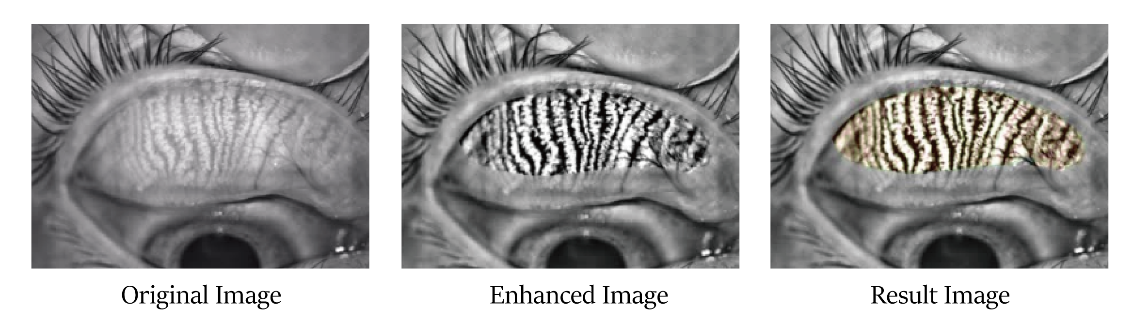
● Meibomian Glands Function Evaluation
● Conjunctival Redness Analysis
● Lipid Layer Thickness
● Blink Quality
● Non-Invasive Tear Film Breakup Time
● Cornea Sodium Fluorescein Staining
● Non-Invasive Tear Meniscus Height
● Eyelid Margin
● Fluorescein Tear Film Breakup Time
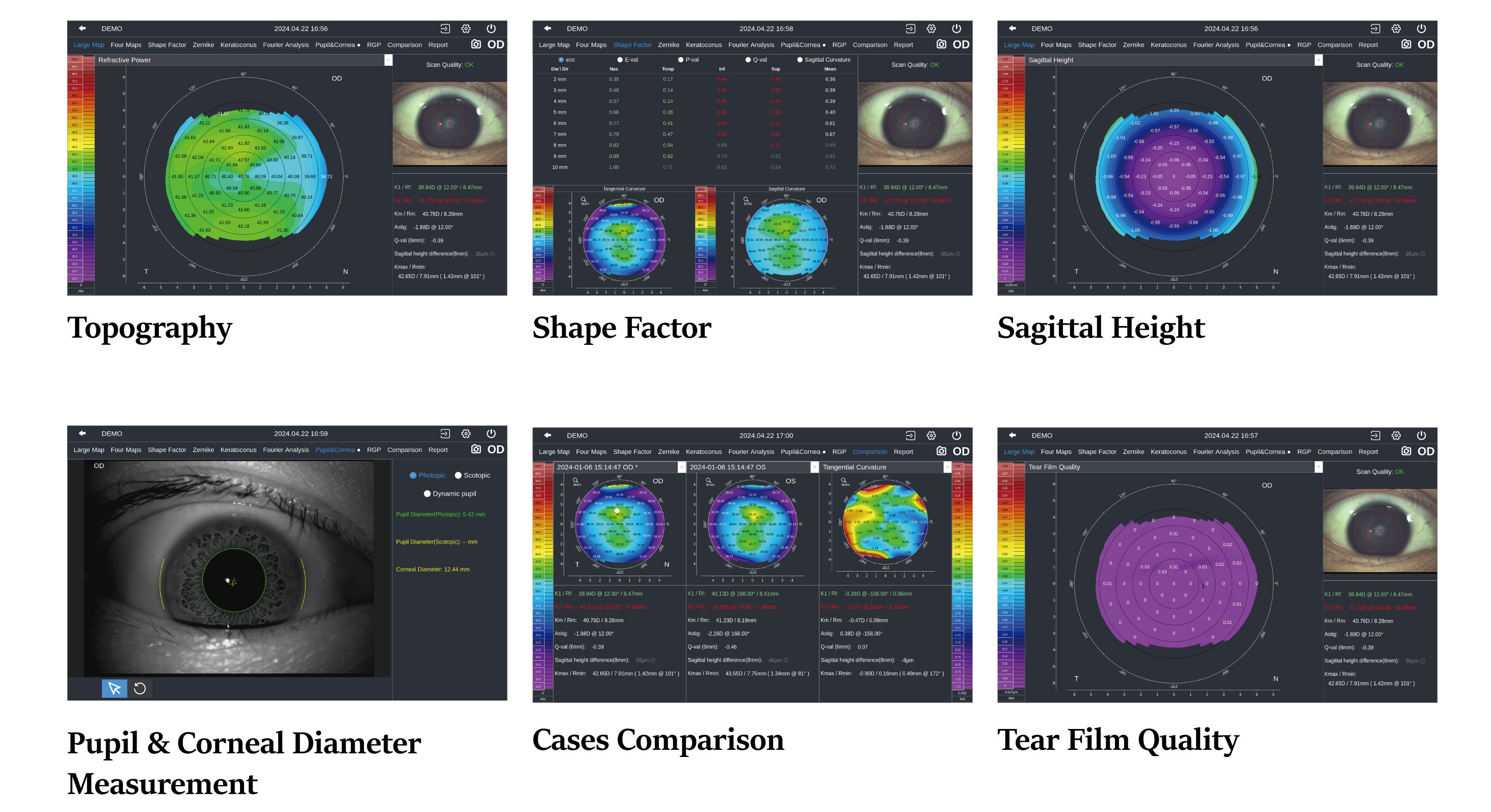
● Topography Analysis
● Aberration & Simulation
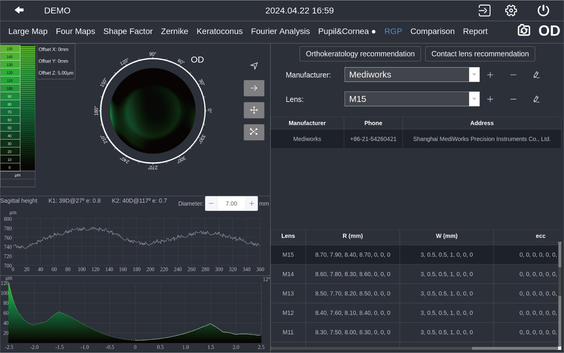
● Lens Fitting
● Pupil & Corneal Diameter Measurement
Integration design enables maximum treatment room utilization
Dry eye diagnosis and Topography analysis integrated
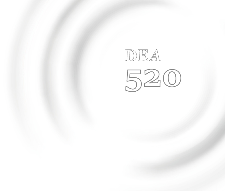
Visualized diagnosis report, easy to understand
External display connection enables real-time observation
Ergonomic Design
50° adjustable display, easier operation.
Auto eyes recognition,switch illumination and magnification intelligently under various function modes.
Compact cone, specially designed for various orbits.
Clinical Application
Dry Eye Analysis
Cornea Morphology Diagnosis
Aberration & Simulation
Lens Fitting
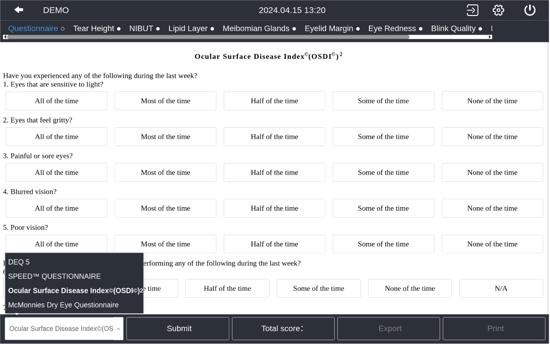
Ocular Surface Disease Index (OSDI)/McMonnies/SPEED/DEQ 5
The built-in dry eye questionnaire is designed according to the risk factors and clinical characteristics of dry eye, providing a simple preliminary assessment for dry eye, improving diagnosis and treatment efficiency and facilitating patient follow-up.
Interface
Comprehensive dry eye examinations.
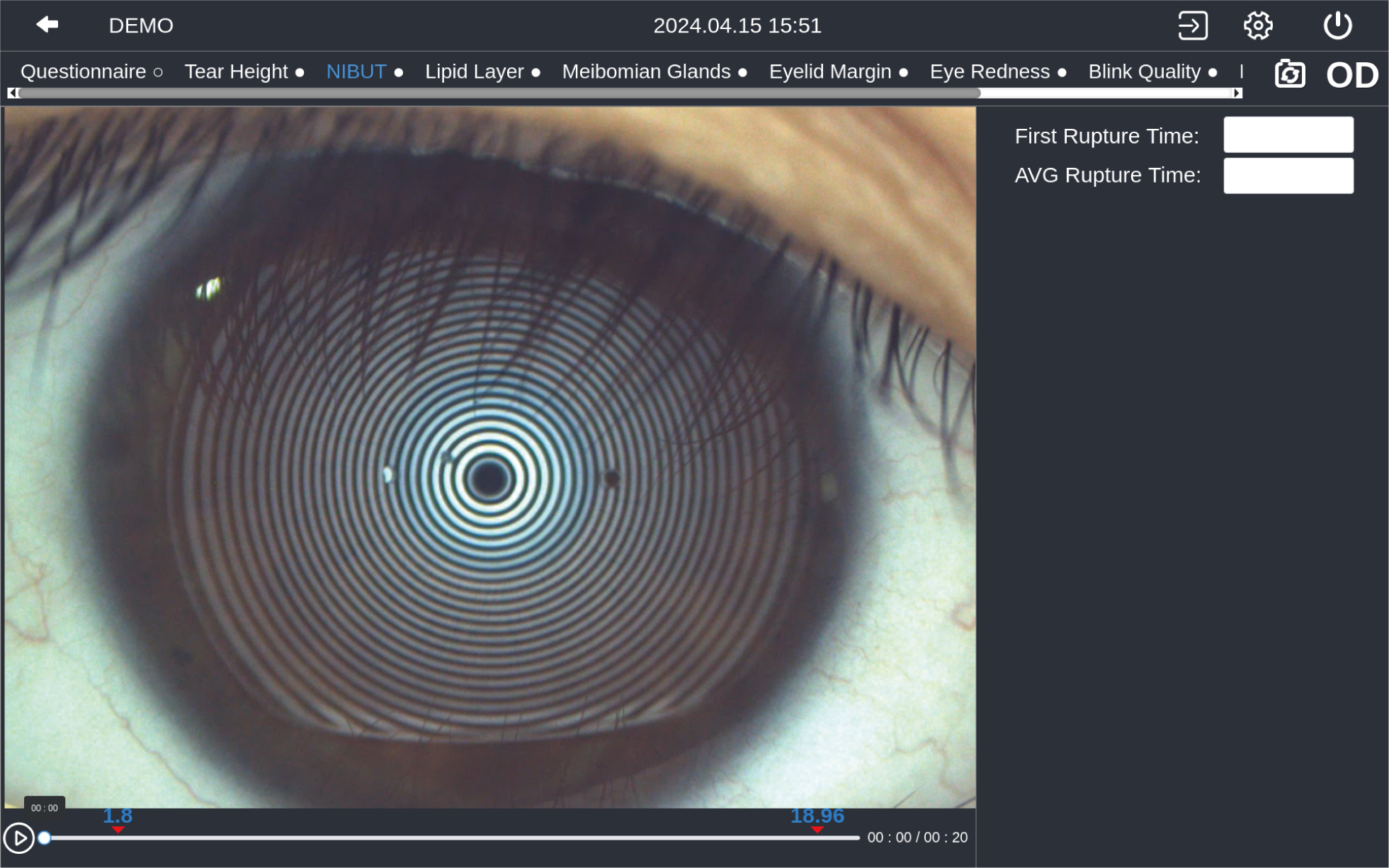
NIBUT
More than 9.6mm diameter Placido ring projection. Auto identify breakup area and analyze NIBUT intelligently.
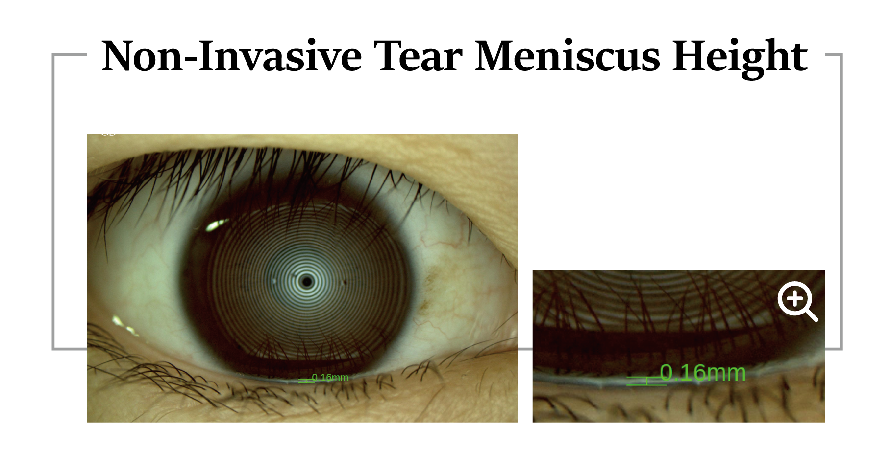
Automatic identification system depicts tear meniscus area and measures the tear height intelligently.
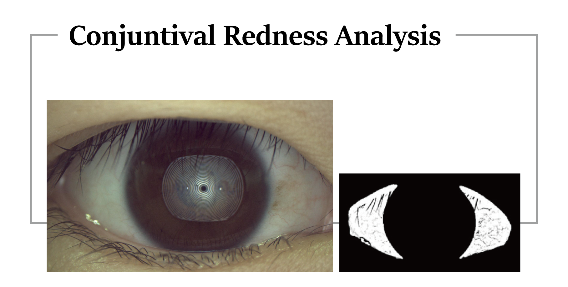
Identify and calculate percentages of conjunctival congestion and ciliary congestions and evaluate severity of eye congestion.
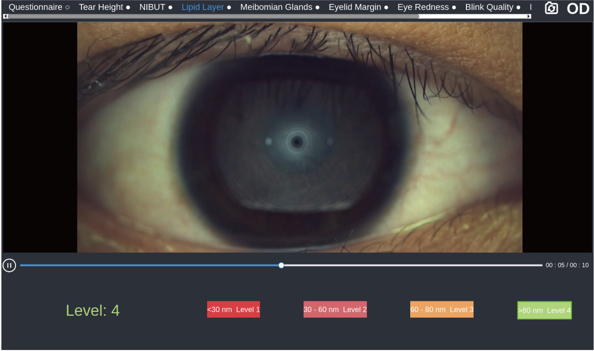
Observe dynamic lipid layer and distribution by video recording compared with standard templates. It’s helpful for judging MGD.
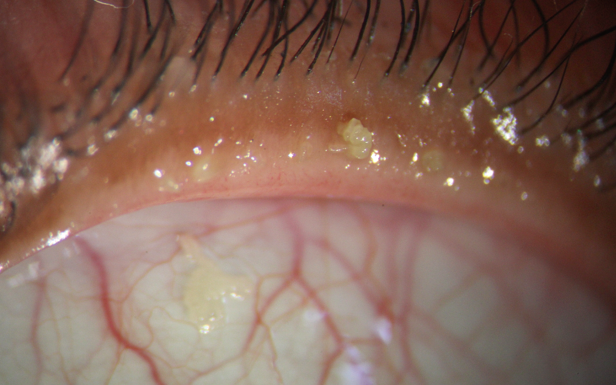
The high resolution image supports zoom in to meet examination requirements of overall shape of eyelid margin and its slight change.
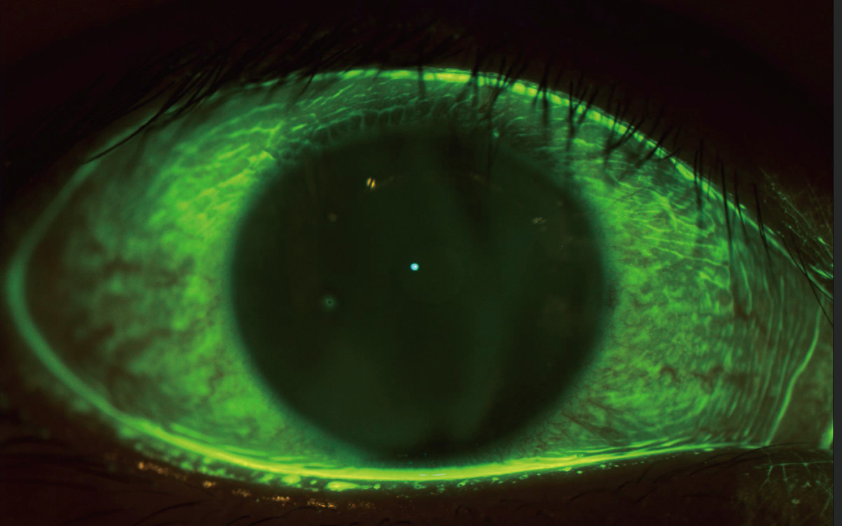
Specially designed built-in yellow filter, working with cobalt-blue illumination improves contrast of corneal fluorescein staining images. Effectively increases positive rate of early corneal epithelial staining.
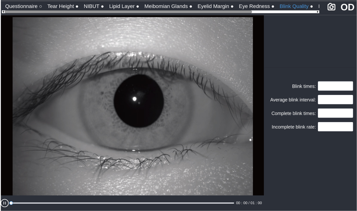
The high-definition video is captured by the infrared light source and automatically analyzes blink frequency, blink interval, incomplete blink, and incomplete blink ratio.
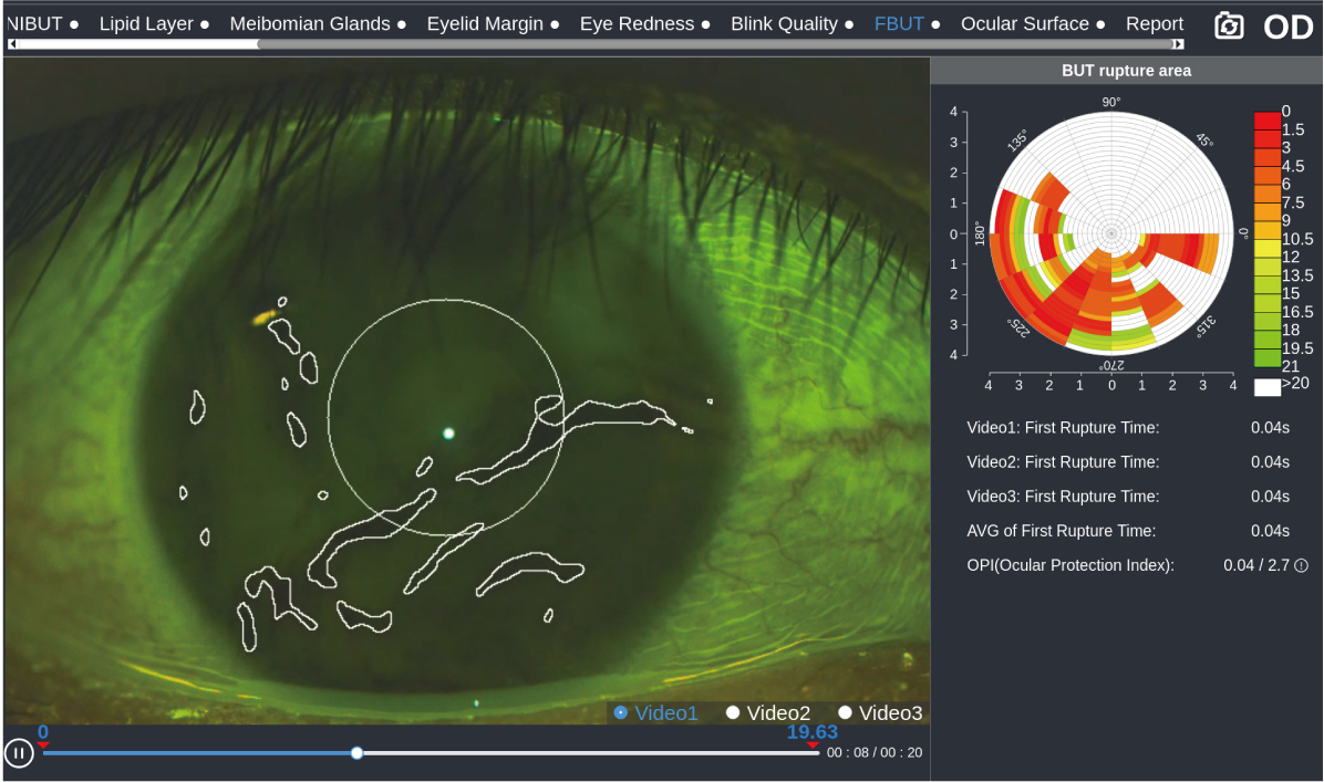
AI automatically detects changes in tear film morphology and calculates tear film breakup time to assess tear film stability.
A simulated fluorescein image will be created based on patient’s cornea. The system will recommend several suitable lens for choose, which accelerates work flow and excludes unfit lens to save the trouble for patient to do real several fluorescein staining.
Research and develop with team SOS from EYE & ENT Hospital of Fudan University. Recommend the most precise lens based on the patient documentation.
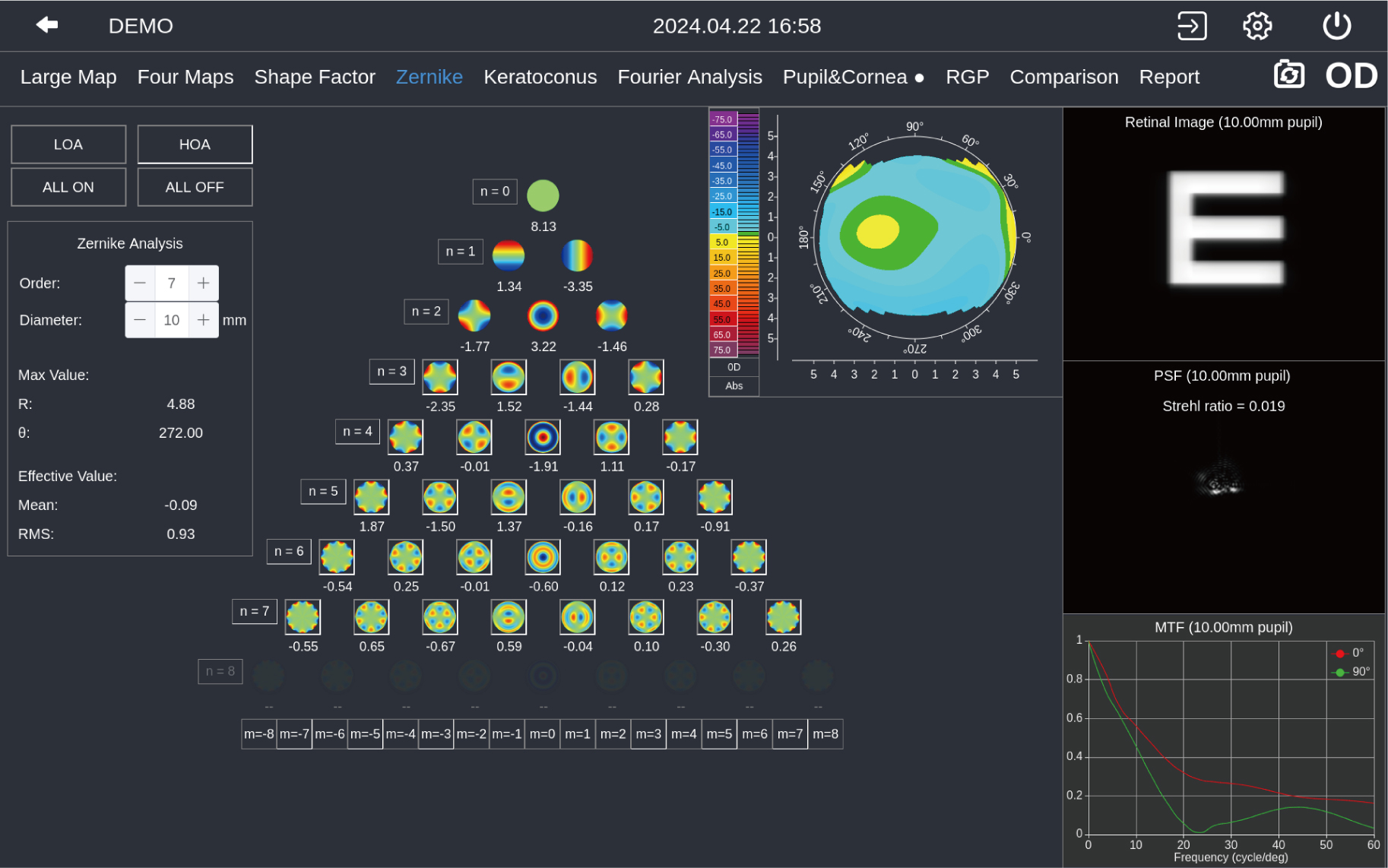
Zernike wavefront aberration analysis makes plan of cataract and refractive surgeries visualized and ensures patient’s postoperative vision quality.
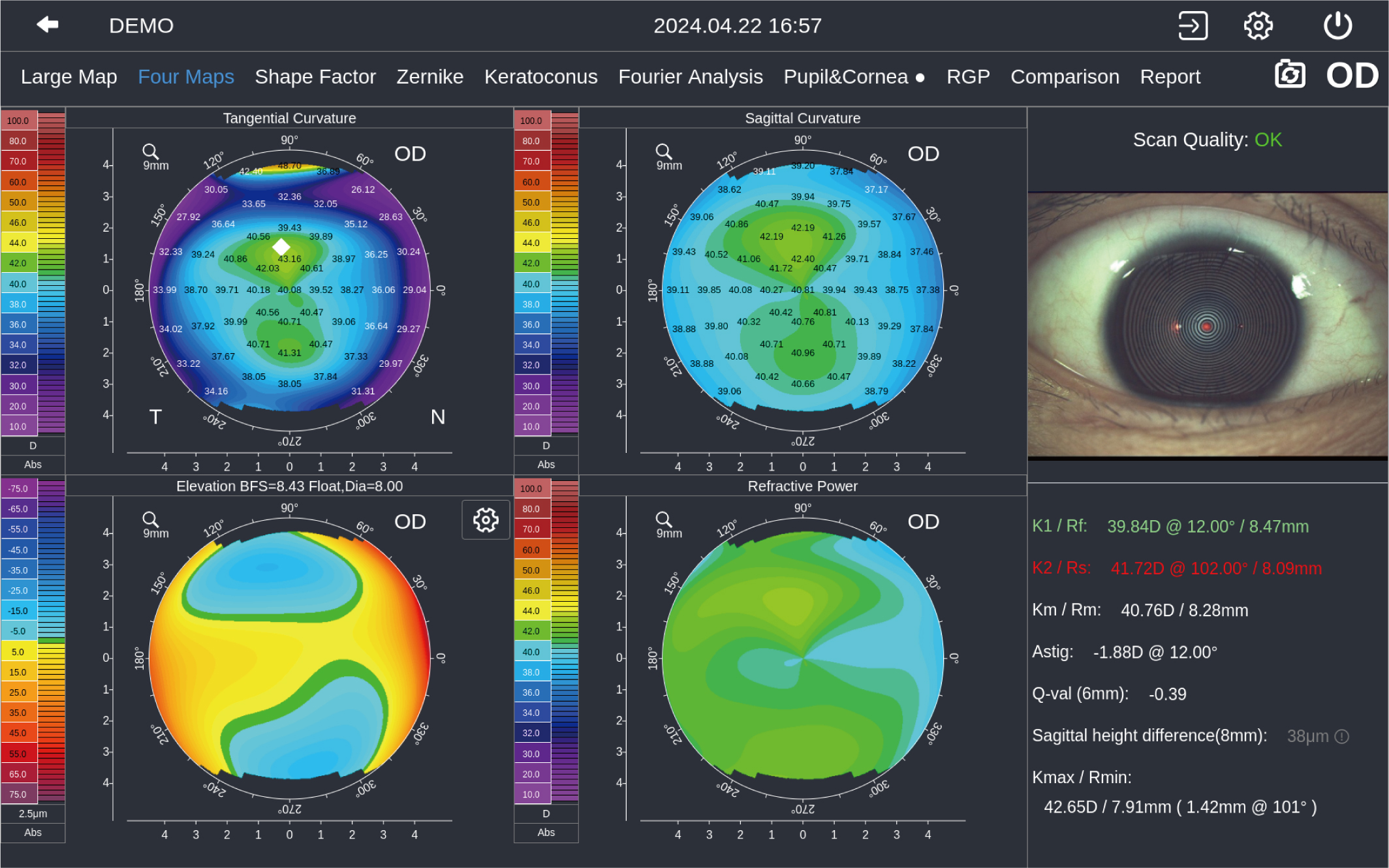
4 maps provide Sagittal Curvature,Tangential Curvature, Elevation Map, Refractive Power, and K1/K2/Km/Astig/Ecc value.
我11
| Dimension | 49 cm × 31 cm × 54 cm |
| Weight | 16.1 kg |
| Built-in CPU | intel |
| Hard Disk | 1 TB |
| Image Resolution | 2048 × 1536 |
| Display | 10.1 ″ touchscreen |
| Illumination | White, Infrared, Cobalt-blue |
| Internet Connection | Wifi & Wired networ |
| Printer Connection | WIFI, USB |
| Power Supply | 100 ~ 240 VAC,50 / 60 HZ |
| Extension Display Interface | Display Port |
| OS/OD Recognition | Automatic |
| Chin Rest Control | Electrical |
| Left and Right | 0 ~ 90 mm work range |
| Front and Back | 0 ~ 60 mm work range |
| Up and Down | 0 ~ 30 mm work range |
| Language | Chinese / English / Japanese / German / Italian |
| DICOM | Supported |
| Numbers of Rings | 50 Rings | |||||
| Diameter of Project Area | 8.8 mm( 42.18 D ) | 11 mm( 42.18 D ) | ||||
| Radius of Curvature | 32.14 dpt ~ 61.36 dpt ( 5.5 mm ~ 10.5 mm ) | Accuracy : ± 0.1 dpt ( ± 0.02 mm ) | ||||
| Astigmatism Axis | 0 ~ 180° | |||||
| White To White | 1 ~ 20 mm | |||||
| Pupil Diameter | 1 ~ 13 mm | |||||
| Topography Function | Sagittal Curvature | Tangential Curvature | Elevation Map | Refractive Power | Sagittal Height | Tear Film Quality |
| 4 Maps | Four Maps display | |||||
| Shape Factor | Ecc, E, p, Q | |||||
| Zernike | Corneal wavefront aberration,PSF map, MTF curve and Simulated image in different pupil diameters | |||||
| Examination Result Comparison | Support 2 results comparison and difference calculation |
| NIBUT | Automatic analysis, tear film rupture area and trend, first break-up time and average break-up time |
| Tear Meniscus Height | 0.01 ~ 2 mm |
| Meibomian Glands | Meibomian glands loss rate and grade |
| Lipid Layer | Template match |
| Eye Redness | Conjuntival congestion percentage |
| Eyelid Margin | Support digital images zoom in |
| Ocular Surface | Built-in yellow filter, Blink Quality, Fluorescein Tear Film Breakup Time |