Application of MediWorks S390L Slit Lamp Microscope in the fitting of orthokeratology lens
1. Ocular surface health assessment
Before fitting orthokeratology lens, it is necessary to examine the patient's anterior segment by slit lamp microscope, observe whether there is active corneal infection or other anterior segment inflammation, evaluate dry eye conditions such as tear quality, and preliminarily determine whether it is suitable for wearing orthokeratology lens.

2. Dynamic and static fitness evaluation
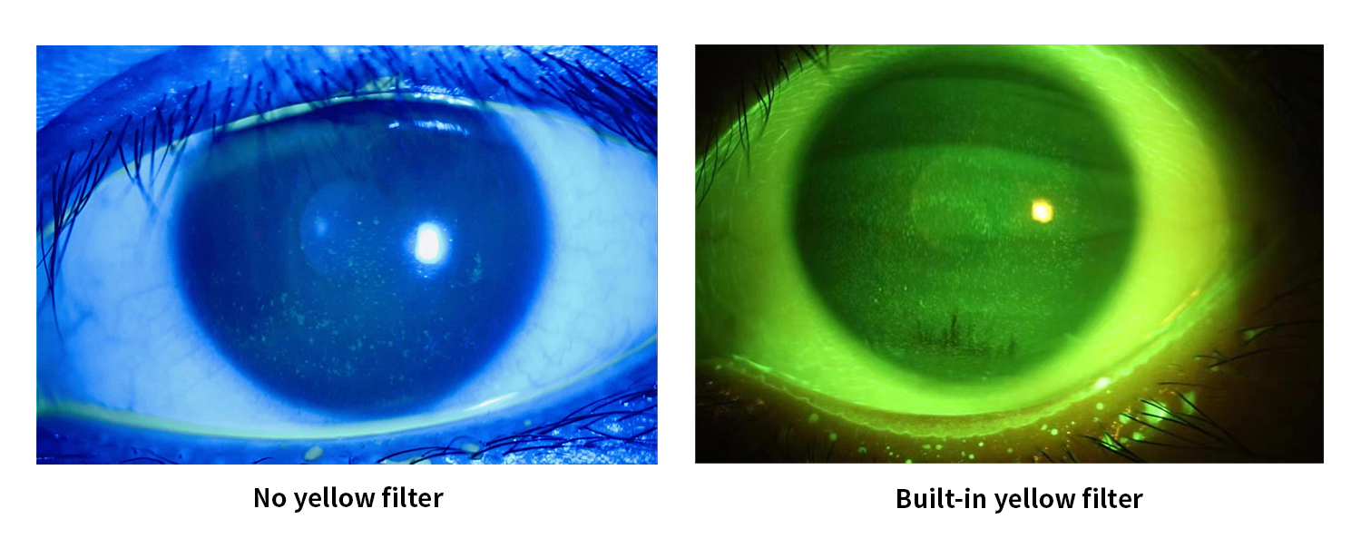
The evaluation of fluorescence matching needs to combine dynamic and static fiting, and pay attention to the central positioning of the lens, the size of the central treatment area, the activity of the lens and the tightness of each curved area.
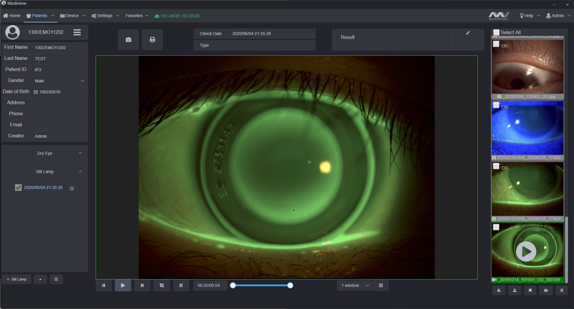
MediWorks S390L Slit Lamp Microscope collects high-definition corneal fluorescein staining photos and blink videos, and built-in yellow filter to increase the contrast of fluorescein staining images.
3. Follow-up evaluation of wearing lens
①Through the slit lamp microscope to observe the patient's eye surface condition, lens adaptation, determine whether it is necessary to adjust the lens parameters. S390L Slit Lamp Microscope supports multi-image comparison function, and the doctor can completely record the process of each lens deployment, which is convenient for communication and follow-up consultation.
② Check the smoothness and integrity of the keratoconus lens surface to determine if there are scratches, protein deposits and aging.
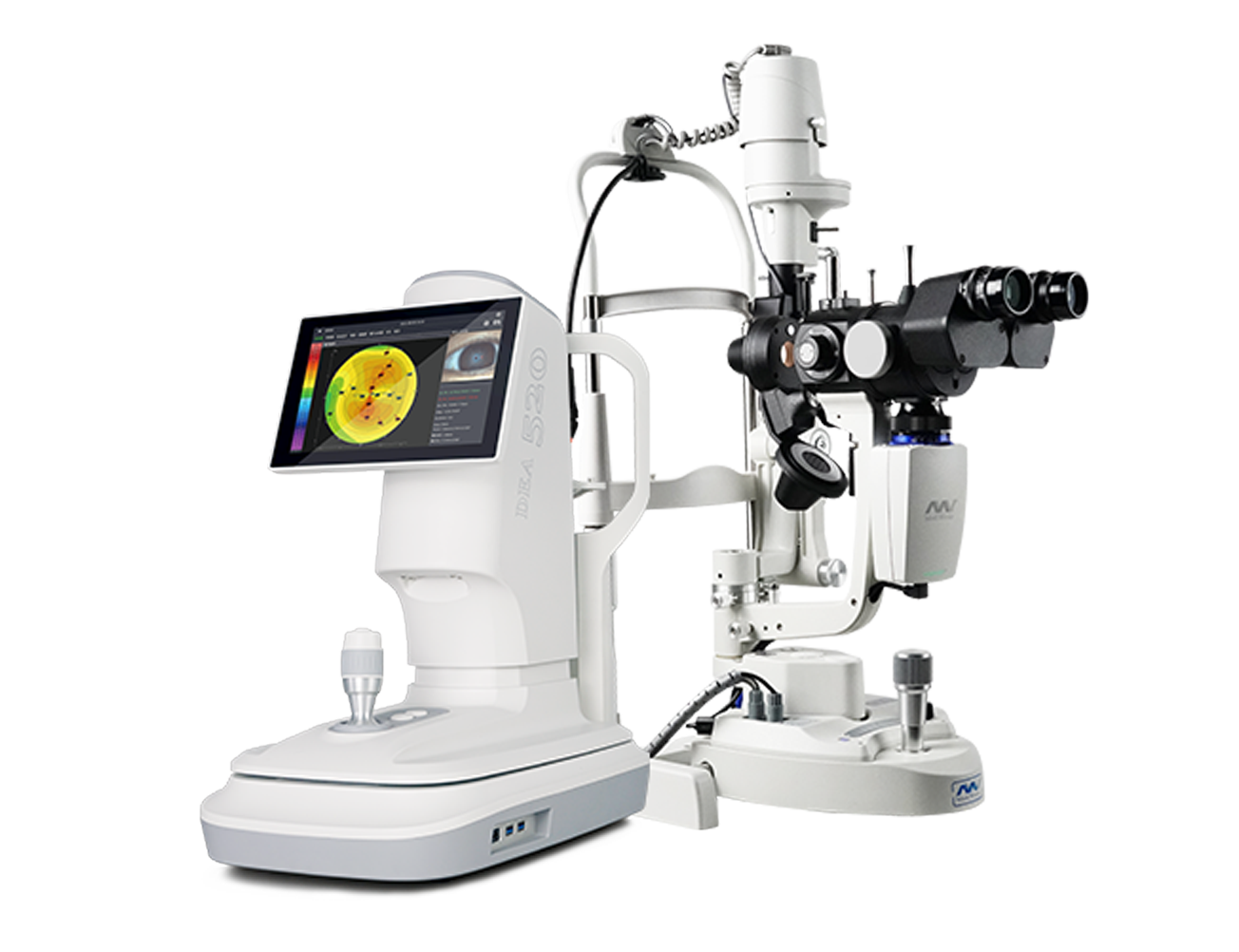
Application of MediWorks DEA520 Corneal Topography in the fitting of orthokeratology lens
1. Before wearing orthokeratology lens:
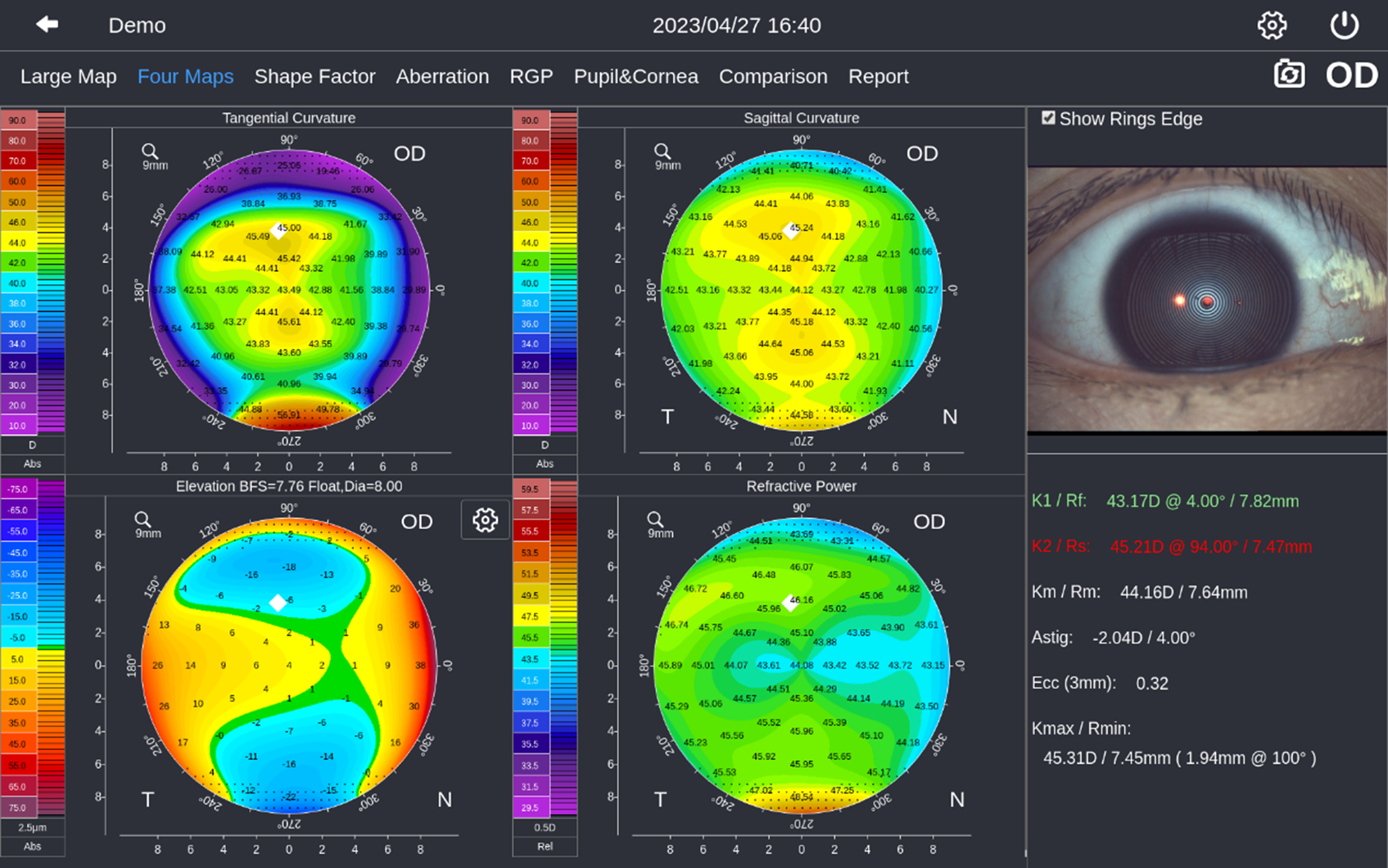
① Quantitative analysis of corneal surface morphology and curvature changes, analysis of corneal astigmatism, and exclusion of abnormal corneal disease.
② The patient's corneal curvature, Ecc value, corneal diameter and other parameters are used to select the appropriate orthokeratology lenses.
2. After wearing orthokeratology lens:
Visually reflect the shape of the cornea after shaping, evaluate the fitting effect, and determine whether the lenses are misaligned, loose or tight, and other undesirable conditions.
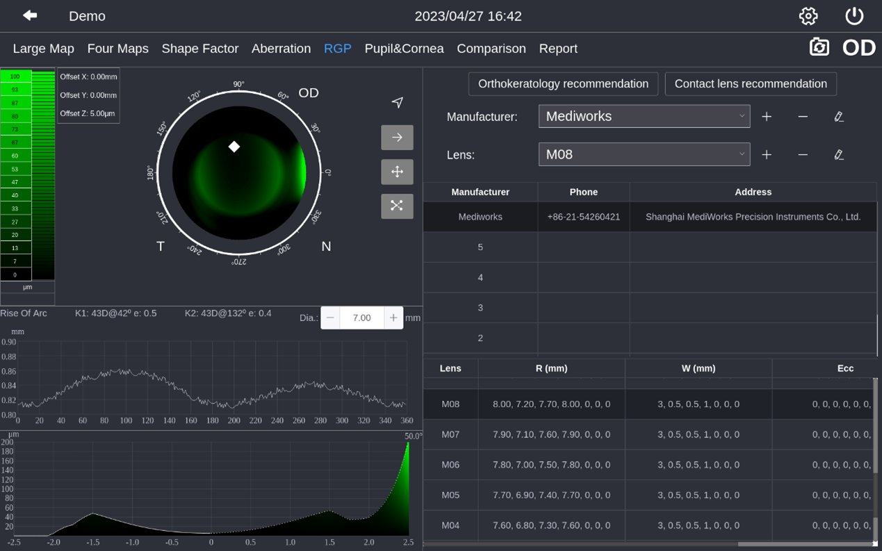
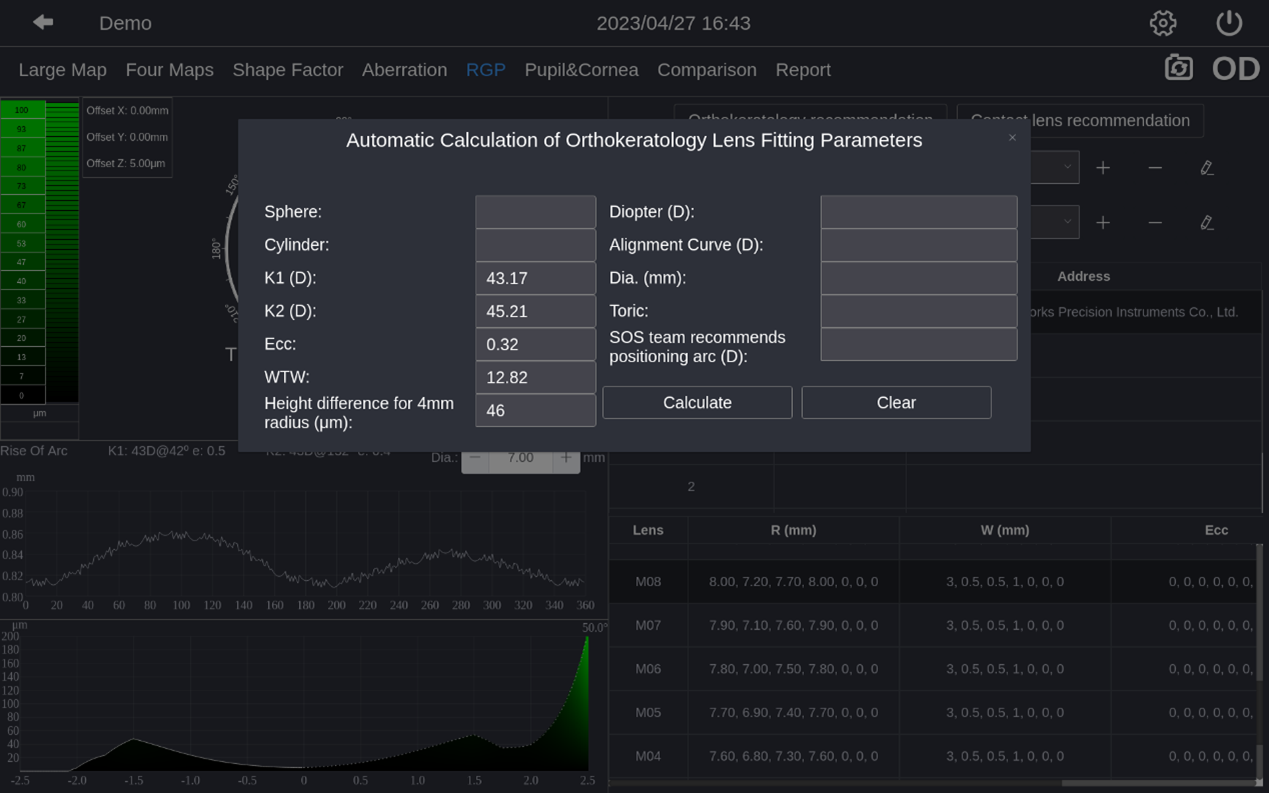
MediWorks DEA520 Corneal Topography supports a custom database to simulate fluorescence staining to improve the fitting efficiency of orthokeratology lenses, eliminating the need for patients to try -on multiple times; it can also automatically recommend appropriate orthokeratology lens fitting parameters based on the patient's corneal morphology.