Doctors usually check the fundus by using relevant instruments to accurately diagnose and treat eye diseases according to the different symptoms shown in the photos shot by the instruments. A fundus camera is one of the instruments to be used. It is a kind of retinal camera allowing ophthalmologists to view the retina more deeply and store the results for further research and comparison. More importantly, doctors and patients can view more explicit images for more in-depth disease analysis and further treatment.
Fundus photography is a process of continuous photography of the eye through the pupil. In ophthalmic medicine, the fundus camera can be divided into a non-mydriatic fundus camera and a mydriatic fundus camera. Which is better for ophthalmologists? The rest of the article will focus on it.
The pupil is shrinking under the intense light, and the traditional fundus camera cannot take a clear and comprehensive picture of the fundus. Therefore, the ophthalmologists will ask the patient to use eye drops before taking photos with mydriatic fundus cameras to increase the size of the pupil for a more evident inner surface of the eye to check.
The use of a non-mydriatic fundus camera means that high-definition pictures of optic disc, retina, and lens can also be achieved by the particular low power microscope of the instrument without increasing the size of the pupil.
Compared with the mydriasis fundus camera, one of the most significant advantages of popularizing the non-mydriasis fundus camera among doctors is its revolutionary upgrade that allows bigger and clearer pictures to take without pupil dilation. It not only is patient-friendly, eliminating the 30-minute waiting time for pupil dilation and eye adjustment time after blinking, but also helps ophthalmologists improve the efficiency of diagnosis.
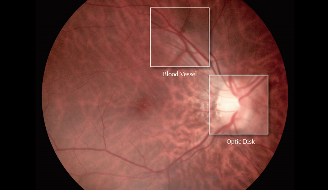
Today's non-mydriatic fundus camera is an easy-to-use revolutionary device that captures detailed images that maintain eye health and contribute to diagnosing eye problems at an early stage. Fundus photography, by the reflective properties of the retina, is good at capturing the subtle details of a variety of severe eye conditions, such as macular degeneration, glaucoma, hypertension, multiple sclerosis, diabetic retinopathy, and so on.
A mydriasis fundus camera is neither superior to non-mydriatic fundus camera in the term of entirety and clarity nor in wide-angle and three-dimensional images to take. Therefore, to establish enough trust and authority in patients, ophthalmologists are more willing to choose a non-mydriatic fundus camera as their best assistant for comprehensive fundus assessment.
Fundus examination plays a vital role in the comprehensive evaluation of a variety of nervous system diseases. Some medical academic research centers can benefit from the purchase of non-mydriatic fundus cameras because a single photo makes it easier to assess and share challenging cases with other clinicians or train the next generation of healthcare professionals, which is of considerable value in large healthcare providers.
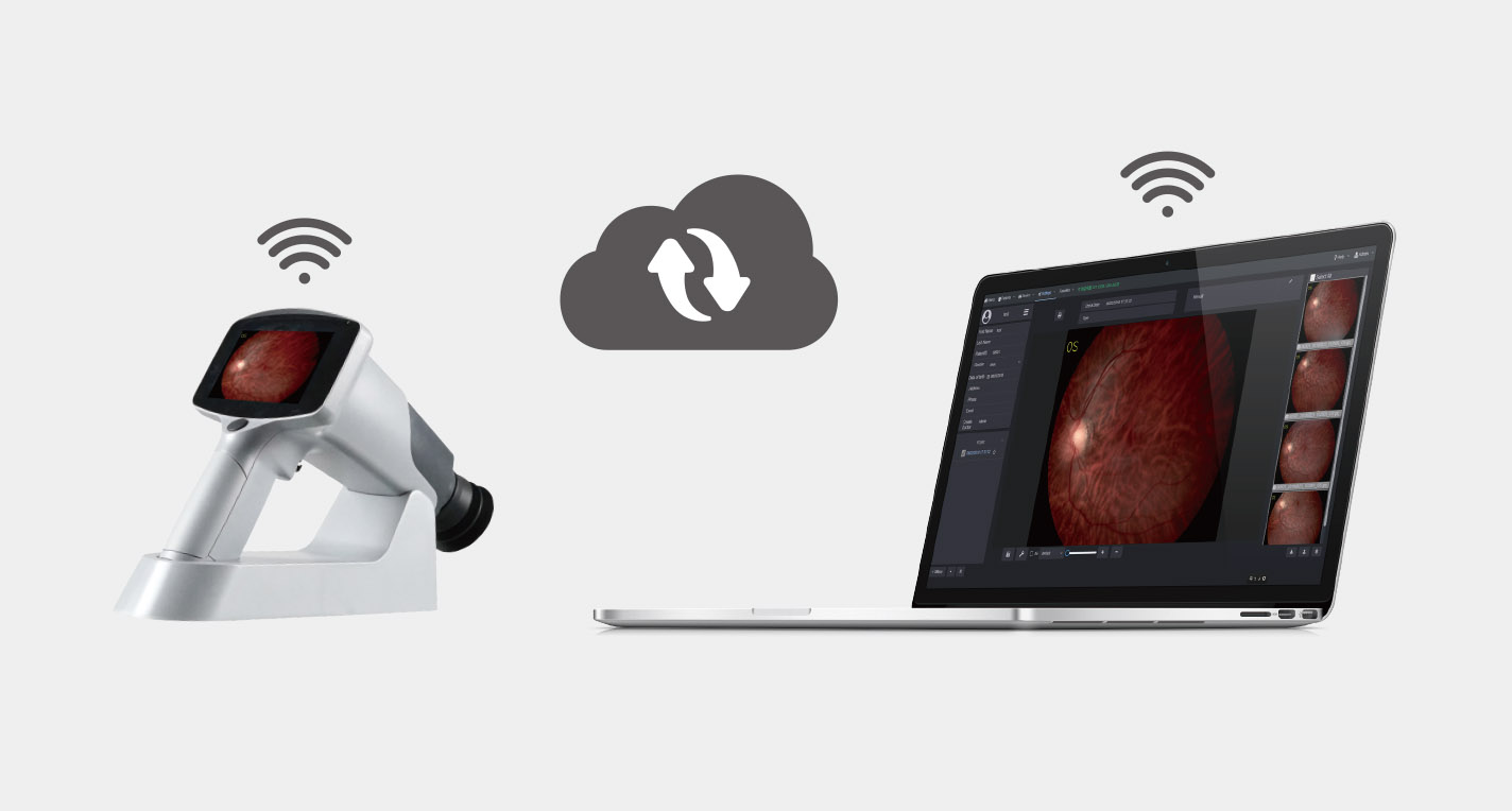
The development of information technology gives ophthalmologists opportunities to realize remote consultation, including the interpretation of fundus photography. By sharing the photos taken by the non-mydriatic fundus camera in the cloud, ophthalmologists from different places can quickly interpret the fundus images and make triage decisions simultaneously.
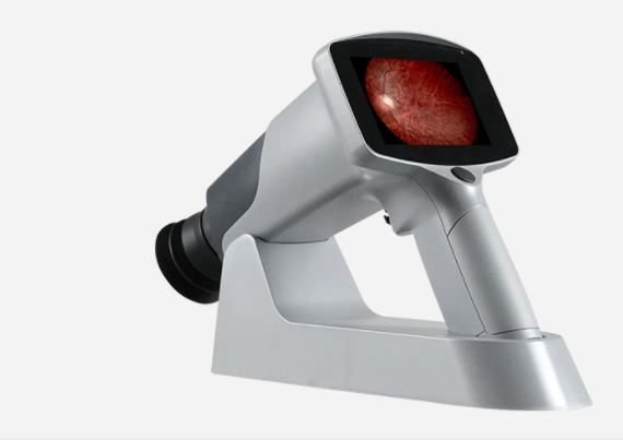
Mediworks FC161 non-mydriatic fundus camera supports real-time synchronization of fundus images between Mediview patient management system through WiFi connection, which is a valuable additional function to carry out cloud sharing and online diagnosis at any time.
Sometimes, it is difficult for doctors to observe optic disc edema using ordinary instruments directly, but a non-mydriatic fundus camera may help. Although non-mydriatic fundus photography may not be able to determine whether the patient has optic disc edema, it can help to find other fundus signs of optic disc edema, including blurred optic disc edge, optic nerve cup filling, folds around the nipple, optic nerve nipple congestion, and venous congestion, etc.
The images obtained by the non-mydriatic camera can detect eye diseases in the early stage so as to start preventive treatment as soon as possible. At the same time, it provides ophthalmologists with more valuable information about the progress of eye diseases and promotes the progress of telemedicine. There are no reasons not to equip a fundus camera in your medical institution. Mediworks non-mydriatic fundus camera is worth trying for its high-resolution fundus photography, 85 ° field of vision covering the fundus, and smart touch LED display bringing more intelligent operation experience to ophthalmologists.