With the advancement of technology, many things change dramatically. For example, the past few years have witnessed a common social phenomenon that an expanding number of people now wear glasses or have some problems in eyes. It is imperative for them to have an examination of eyesight and optometry regularly. However, the subjective examination in optometry mainly relies on the patient’s statement and feeling like the past judgment criterion. There are inevitably some differences between disparate people so that the examination results fluctuate considerably. Therefore, a professional anterior segment analyzer is of great importance.
In the front part of the eye, the Anterior Segment Analyzer measures almost everything. You may or may not be able to see far through the angle or behind the iris, depending on the wavelength, so that you can see the different corneal layers in detail, and there are ways to look at the anterior and posterior curvatures of the cornea. For that purpose, when performing different types of corneal surgery, this technology has great value for an anterior segment surgeon, especially in corneal pathology cases or as a pre-or postoperative method.
Anterior segment analyzer imaging system in clinical application for better analysis of the disease and providing professional solutions to clinicians have valuable values; it is used for several purposes:
When having LASIK done, anterior segment analyzer is beneficial in determining the quality of the corneal surface and can distinguish an existing LASIK flap from the surrounding tissue.
It is a sensitive test for diagnosing forme fruste keratoconus when determining if a patient is suitable for LASIK.During cataract surgery, anterior segment analyzer can help measure the anterior chamber angle area, which aids in better cataract surgery planning for anterior segment surgeons.
Additionally, it also provides professional data of total corneal astigmatism, total spherical aberration and corneal membrane irregularity during cataract surgery, giving analysis support for solving spherical lens ametropia astigmatism, spherical aberration, and presbyopia.
The scansys anterior segment analyzer can provide the prevalence of keratoconus by using the AI algorithm and Further checking the topographic maps to accurately analyze and diagnose the keratoconus. And finally, according to the presence of keratoconus to determine whether keratoplasty can be performed.
Using a scansys anterior segment analyzer to examine the situation of eyes is quite essential before ICL lens surgery. It can accurately analyze the changes in the morphology and curvature of the entire corneal surface and systematically and objectively analyze the examination of corneal characteristics.
The anterior segment analyzer supports disparate angles to collect a high-definition picture to provide adequate data support for ICL surgery. The corneal curvature will be finally shown in data and different colors clearly. Therefore, the success rate of the surgery has also significantly increased.
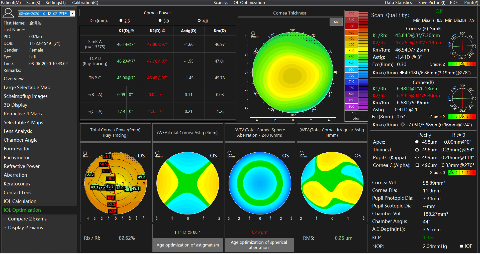
It is specially designed for the “IOL Optimization” of refractive cataract surgery. The scansys anterior segment analyzer provides special reference values such as corneal refractive type.
It is worth mentioning that the scansys anterior segment analyzer provides professional data of the total corneal astigmatism aberration, total corneal spherical aberration, and the total corneal irregular astigmatism, and analysis support for solving spherical refractive errors, astigmatism, spherical aberration, and presbyopia in cataract surgery.
The scansys anterior segment analyzer provides a key parameter column obtained in the range of 3mm in diameter of the membrane. In order to describe in more detail the difference of these values in each diameter range, true net refractive power, whole cornea refractive power can be seen in the corneal topography machine.
Furthermore, the changes of these values in different topographic maps and different diameter ranges are described more intuitively and in detail. Finally, total cornea aberration guides surgeons to evaluate preoperative and postoperative visual quality to ensure patients of best surgery effect.
It's worth wondering why it's catching on too slowly, not that not useful for the anterior section. The truth is that surgeons can get pictures and extract a lot of valuable details from them; thus, grabbing an anterior segment analyzer is worthy. Here at Mediworks’s Scansys TA517 is the right hand for anterior segment surgeons for AI Keratoconus diagnosis, IOL optimization, ICL surgery examination, and aberration analysis. With the increasing interest in assessing dynamic factors such as changes in iris area or volume as only static factors were focused on before, Mediworks’s Scansys TA517 must play an important role in further diagnosis.