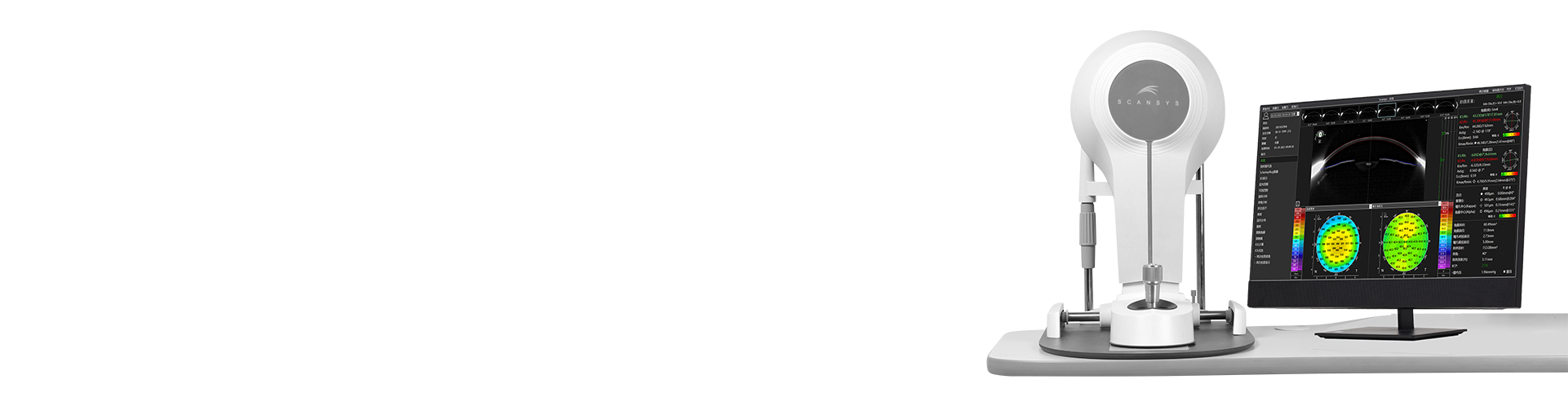
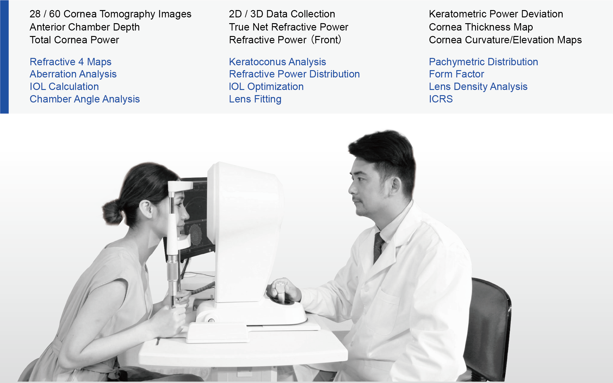
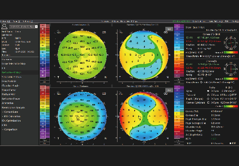
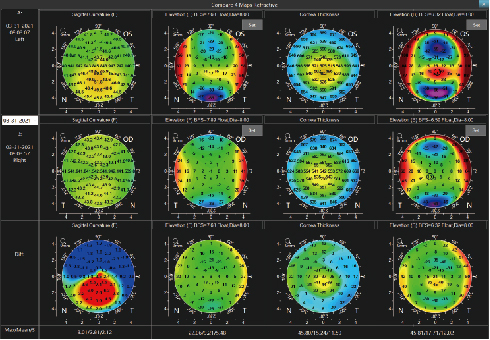
Scansys provides a professional solution for anterior segment diagnosis. It applies Scheimpflug camera which can collect 107520 / 230400 data points and generates 28 / 60 cornea tomography images in high resolution. Scansys can provide a series of topographic maps including cornea curvature maps, cornea thickness maps, cornea elevation maps, etc.
◉Keratoconus Diagnosis
Scansys can provide the prevalence of Keratoconus by using the algorithm, Further checking the topographic maps to accurately analyze and diagnose the keratoconus.
◉ICL Surgery Examination
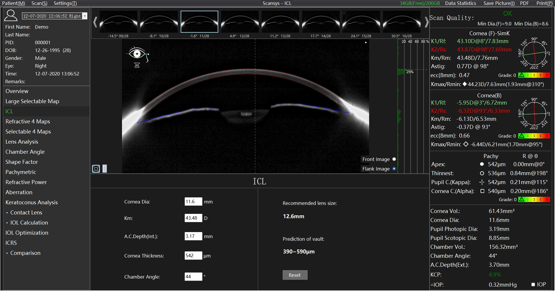
Scansys supports to collect a high-resolution Scheimpflug images in different angles. It also provides White to White, AC depth for ICL surgery.
Artificial intelligence recommends the diameter of the ICL and gives the prediction of postoperative vault.
◉IOL Optimization
Specially designed for cataract surgery. It supports clinicians to choose suitable Toric IOL, Aspheric IOL or Multifocal IOL for patients.
◉Aberration Analysis
Total cornea aberration guides surgeons to evaluate preoperative and postoperative visual quality to ensure the best surgery effect for patients.
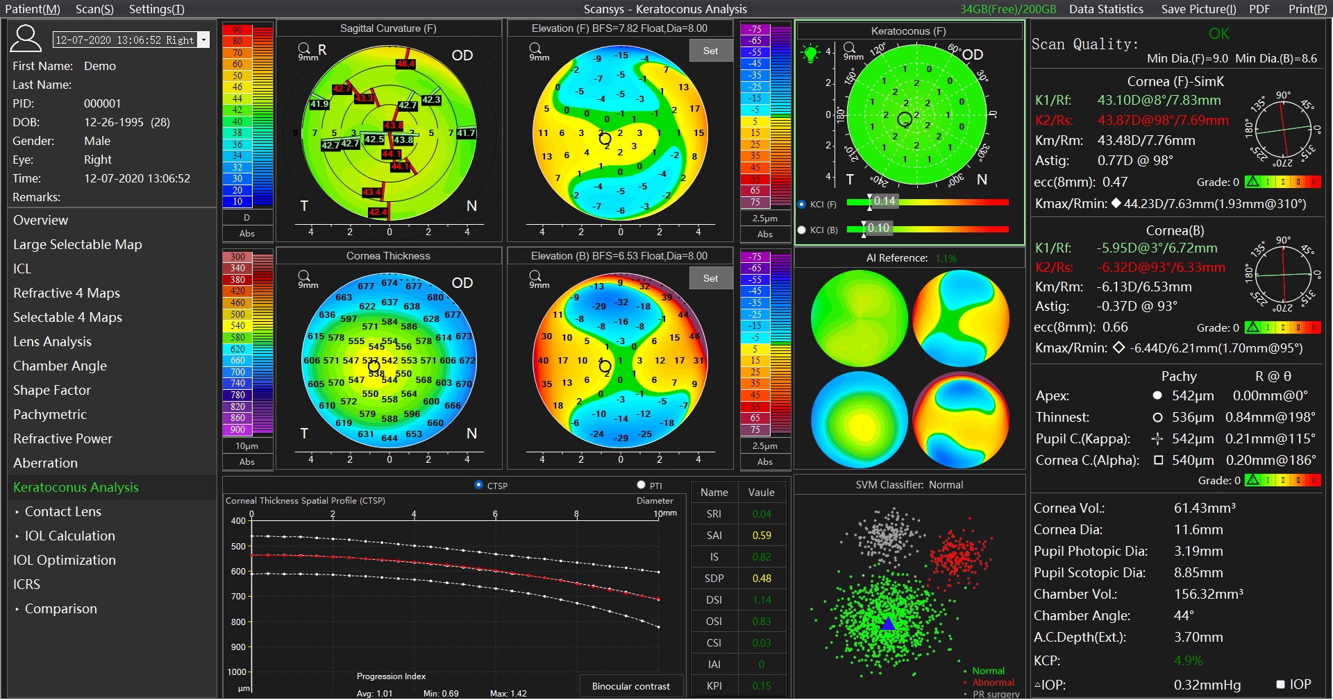
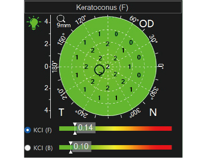
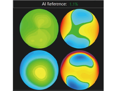
Keratoconus analysis module gives the KCI index , reference, SVM classifier, keratoconus possibility(KCP), refractive 4 maps, corneal thickness spatial profile (CTSP), percentage thickness increase (PTI) and so on to help doctors quickly and intelligently screen keratoconus.
abnormal protrusion index of anterior and posterior corneal surface.
◆ Color in green indicates the cornea is normal.
◆ Color in yellow indicates a suspicious abnormal protrusion of the cornea.
◆ Color in red indicates an abnormal protrusion of the cornea.
Reference introduces algorithms to more intelligently screen keratoconus.
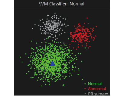
After learning the characteristic points of different cases,
Scansys classifies the current cases.
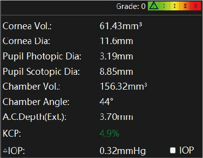
Values are distributed between 0-100%, the larger the value, the greater the probability of keratoconus. The unique KCP parameter is used to intuitively judge the possibility of keratoconus.
Sagittal Curvature (Front), Corneal Thickness, Elevation (Front), Elevation (Back)
It’s a key reference for specifically diagnosing the keratoconus.
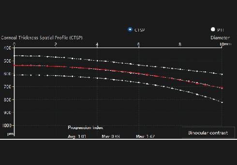
The trend distribution of the corneal thickness.
It is efficient for the early detection of keratoconus and corneal ectasia and improves the sensitivity and specificity of early keratoconus and ectasia screening.
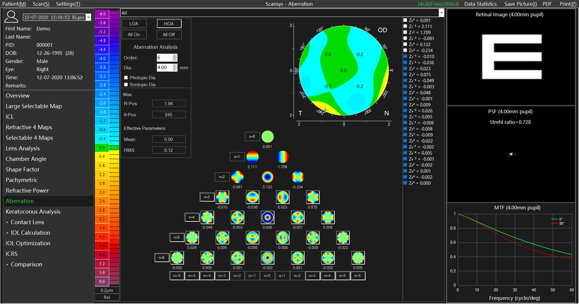
This module analyzes the aberrations of the anterior surface, posterior surface of the cornea and the total cornea through the zernike polynomial and provides accurate information for optical correction through retinal image, point spread function, modulation transfer function, which has guiding significance for the visual quality analysis before and after refractive surgery.
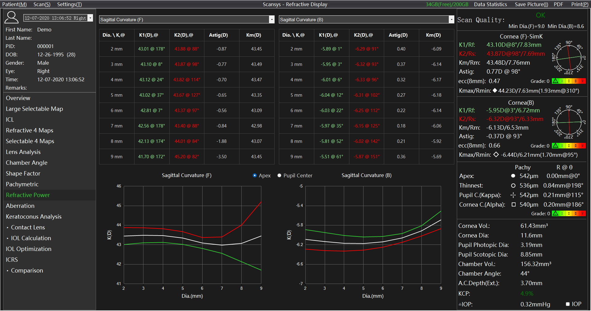
In the key parameter column on the right, we give K1, K2, Km, Astig. These values are obtained in the range of 3mm in diameter of the membrane. In order to describe in more detail the difference of these values in each diameter range, We give K1, K2, Km, Astig of the axial curvature of the anterior and posterior cornea, anterior surface refractive power, true net refractive power, full corneal refractive power topographic map, the distribution table of various areas from 2mm to 9mm in diameter and the distribution curve .The changes in these values in different topographic maps and different diameter ranges are described more intuitively and in detail.
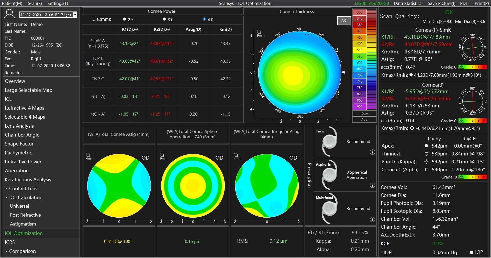
Specially designed for the “IOL Optimization”of refractive cataract surgery. Given the K1, K2, Km, and Astig values of the three types of corneal refractive power (Simk, total corneal power, true net refractive power), and Kappa & Alpha angle, respectively. It also provides professional data of the total corneal astigmatism aberration, total corneal spherical aberration and the total corneal irregular astigmatism,and analysis support for solving spherical refractive errors, astigmatism, spherical aberration, and presbyopia in cataract surgery.
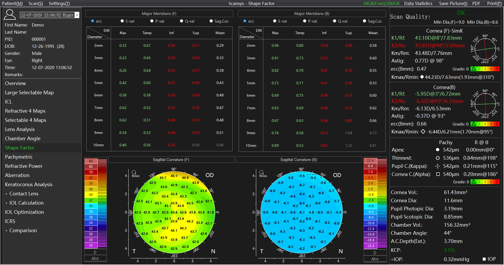
The chart upper depicts the corneal form factor and the curvature of the anterior and posterior surface of the cornea at the intersection of the radial axis and the four radial directions.
Corneal shape factors include Ecc, E, Q, and P, which can be changed in the “Map and Data Settings” -> “Form Factor Presentation” in the menu bar “Settings” options.
↑Preoperative diagnosis & Postoperative arch evaluation
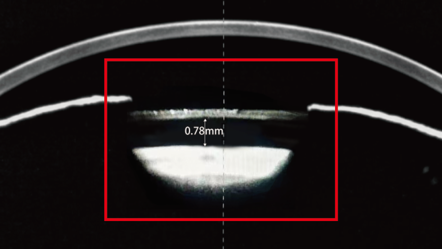
←Postoperative arch measurement
Scansys supports in any Angle to collect a high-definition picture, to provide effective data support for ICL surgery.
Intelligence recommends the diameter of the ICL and gives elevation of the arch.
Scansys calculates the lens density value for cross section and longitudinal section which is helpful in cataract diagnosis.
A simulated fluorescein image will be created based on the patient’s topographic maps generated by Scansys. This will accelerate the work flow of lens fitting and save the trouble for patient to accept multiple real fluorescein staining during lens fitting.
VST-Four arcs and above design
Input R value, W value and K value of each arc of VST lens.
CRT-Three arcs design Input sphere
Scansys and manufacturer recommendations.
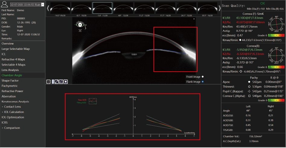
Scansys can calculate a chamber angle value based on the tomography images and its exclusive AOD graph gives a trend analysis for the distance between cornea back surface to iris. It also provides cornea volume, anterior chamber volume and anterior chamber depth calculation. These analyses are used for quickly screening glaucoma.
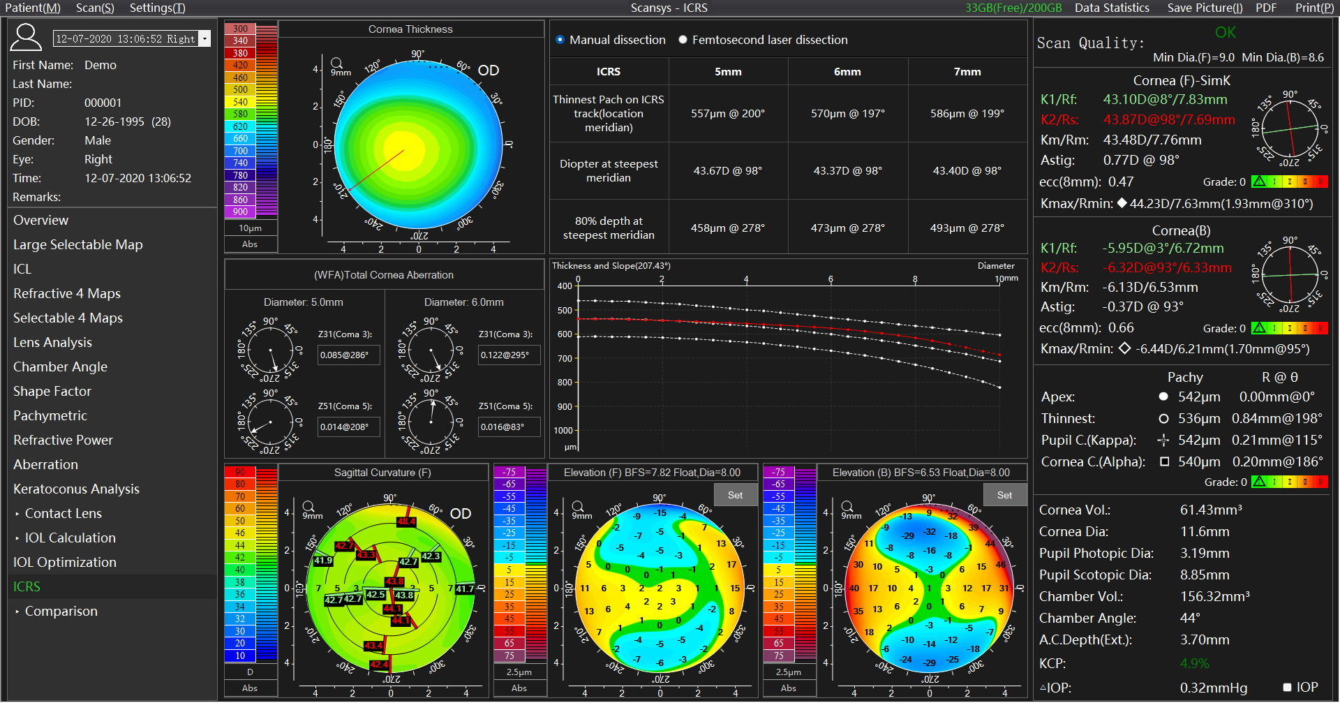
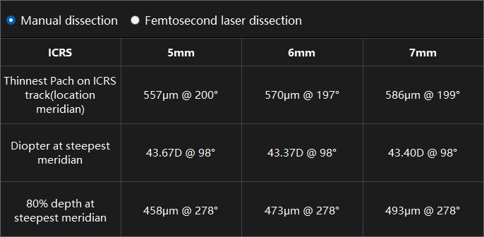
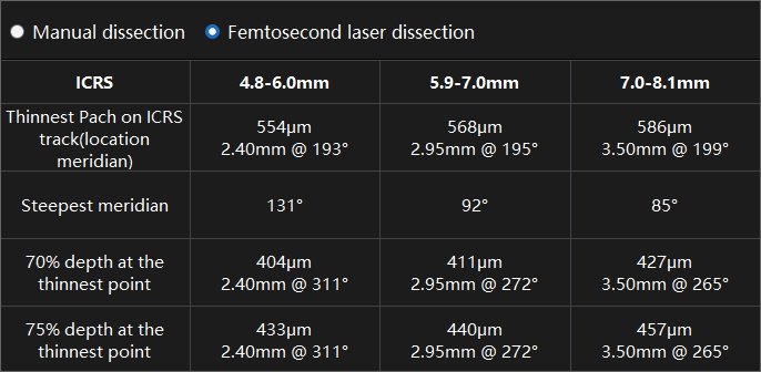
Making a stromal tunnel is the first step in intrastromal corneal ring segments (ICRS) implantation. Based on the patient's corneal morphology, several parameters required to make the stromal tunnel with conventional mechanical (Manual dissection) and femtosecond laser (Femtosecond laser dissection) are provided separately, reducing postoperative complications and improving the predictability of the surgery.
| Camera | Digital infrared camera + Scheimpflug digital CCD camera |
| Light Source | LED slit |
| Scanning Speed | 28 images within 1 second/ 60 images within 2 second/ single image |
| Data Points | 107520 / 230400 |
| Work distance | 80 mm |
| Corneal topography | 9 mm / 12 mm |
| Corneal thickness | 300 ~ 900 μm |
| Anterior chamber depth | 0.8 ~ 6 mm |
| Diopter | 12 ~ 72 D |
| White to white | 6 ~ 14 mm |
| Pupil diameter | 1 ~ 10 mm |
| Anterior chamber volume | 15 ~ 300 mm³ |
| Chamber angle | 16 ~ 60 ° |
| Kappa/Alpha | R( 0 ~ 3 mm) θ( 0 ~ 360° ) |
| Work Range | |
|---|---|
| Front and back | 115 mm |
| Left and right | 100 mm |
| Up and down | 30 mm |
| Power Supply | |
|---|---|
| Input voltage | ~100 V ~ 240 V |
| Input frequency | 50 Hz / 60 Hz |
| Weight and Size | |
|---|---|
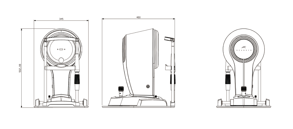
| Device dimension | 505 mm x 345 mm x 460 mm ( L/W/H ) |
| Device weight | 12 kg |
| Package dimension | 700 mm x 600 mm x 830 mm ( L/W/H ) |
| Package weigh | 25 kg |
| System Specifications | |
|---|---|
| PC configuration | i5 ~ 10500T 8GB memory 256GB SSD + 1TB storage |
| Display | 1920 × 1080 23.8 inch |
| PC system | Windows 10 |